- info@careermakers.edu.np
- +977 1 4423870
Biaxin
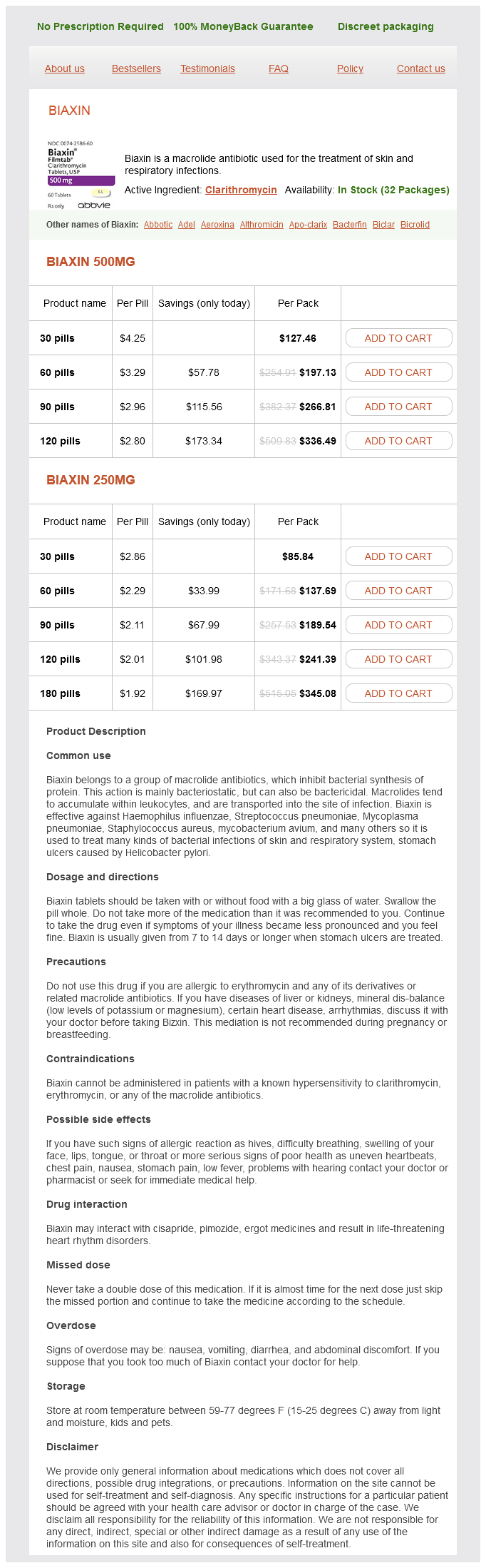
Biaxin 500mg
- 30 pills - $127.46
- 60 pills - $197.13
- 90 pills - $266.81
- 120 pills - $336.49
Biaxin 250mg
- 30 pills - $85.84
- 60 pills - $137.69
- 90 pills - $189.54
- 120 pills - $241.39
- 180 pills - $345.08
Biaxin dosages: 500 mg, 250 mg
Biaxin packs: 30 pills, 60 pills, 90 pills, 120 pills, 180 pills
In stock: 502
Only $2.04 per item
Description
In the long bones of the extremities the shaft or diaphysis is the hard compact portion gastritis duodenitis buy biaxin 500 mg amex, the epiphysis or end is spongelike and covered by a shell or harder bone and the metaphysis or growing portion lies between them. Bone cells multiply rapidly in early years but later on only dead cells are replaced or injured ones are repaired. Long bones give support, flat bones provide protection for delicate organs and irregular bones allow for more motion. The outer portion of bones is hard, however the hollow inner part is filled with soft marrow (Gr. Yellow marrow is found in long bones, whereas red marrow is found in the end of long bones as well as in ribs and bodies of the vertebrae. The latter is responsible for the manufacture of red blood cells, some of the white blood cells and platelets. Bones are usually completely hardened by about twenty years of age through the deposit of calcium and phosphorus from food. Some remain as cartilage, however, for example, the end of the nose, the ears and the anterior part of the ribs attached to the sternum. Where the surfaces of the ends of the bones come together they are covered with a thin layer of cartilage. Inside, the cells of the lining of the joint give out a small amount of slippery fluid (synovial fluid) which keeps them lubricated and allows free movement. Joints have: (1) no movement, for example, the flat bones of the skull; (2) slight movement, for example, the bodies of the vertebrae; (3) free movement, for example, the joints at the shoulder and hip. Ties of freely movable joints (1) Ball and socket A joint in which a rounded head is received into a cup-like socket, for example, the shoulder joint, formed by the head of the humerus and the glenoid cavity of the scapula. Bones of the cranium Two parietal bones, one occipital bone, and one frontal bone form a covering for the brain. Two temporal bones contain the ear cavities, the organs of balance, and the mastoid cells. Two inferior turbinate bones in the nostrils form the outer walls of the nasal cavity. Sinuses Four pairs of cavities in the cranial bones make the skull lighter and return the sound of the voice. Named after the bones in which they lie, there are 2 frontal sinuses, 2 maxillary sinuses, 2 ethmoid sinuses, and 2 sphenoid sinuses. Sinusitis the effect of swollen epithelial tissue which blocks drainage channels and thereby prevents normal secretions in the sinuses 4.
Carica Papaya (Papain). Biaxin.
- Are there safety concerns?
- What other names is Papain known by?
- Dosing considerations for Papain.
- Digestion problems, diarrhea, hayfever, runny nose, psoriasis, cancer, treating infected wounds, sores, ulcers, intestinal worms, and other conditions.
- Sore throat and throat swelling (pharyngitis).
- What is Papain?
- Herpes zoster (shingles).
Source: http://www.rxlist.com/script/main/art.asp?articlekey=96115
They together with osteoclasts play an important role of homeostasis by helping to release calcium gastritis treatment dogs generic biaxin 500 mg otc. They are believed to be derived from osteoblast that ceases their physiological activity. Bone in embryo develops in two ways: Intra-membranous ossification, If bone develops directly from mesenchymal tissue. Endochondrial Ossification, When bone tissue develops by replacing hyaline cartilage. The 64 Human Anatomy and Physiology cartilage it self do not converted into bone but the cartilage is replaced by bone through the process. Endochondrial ossification produces long bones and all other bones not formed by intra-membranous ossification. These are surface markings where muscles, tendons and ligaments attached, blood & lymph vessels and nerves pass. Depression and openings Fissure narrow, cleft like opening between adjacent parts of bone. Example: External auditory meatus Groves and sulcus: are deep furrow on the surface of a bone or other structure. Example Medial condyle of femur Head, expanded, rounded surface at proximal end of a bone often joined to shaft by a narrowed neck. Trochanter: it is a large, blunt projection found only on femur Crest is a prominent ridge. Upper & lower extremities and bones of girdles are grouped under appendicular skeleton. The upper part of the lower extremity, between the pelvis and knee, is the thigh; the leg is between the knees an ankle. Made up of horizontal, cribriform plate, median perpendicular plate, paired lateral masses; contains ethmoidal sinuses, crista galli, superior and middle conchae. Forms roof of nasal cavity and septum, part of cranium floor; site of attachment for membranes covering brain. Shaped like large scoop; frontal squama forms forehead; orbital plate forms roof of orbit; supraorbital ridge forms brow ridge; contains frontal sinuses, supraorbital foramen. Slightly curved plate, With turned- up edges; made up of squamous, base, and two lateral parts; contains foramen magnum, occipital condyles, hypo-glossal canals, atlanto-occipital joint, external occipital crest and protuberance. Protects posterior part of brain; forms foramina for spinal cord and nerves; site of attachment for muscles, ligaments. Parietal (2) Superior sides and roof of cranium, between frontal and occipital bones. Wedge-shaped; made up of body, greater and lesser lateral wings, pterygoid processes; contains sphenoidal sinuses, sella turcica, optic foramen, superior orbital fissure, foramen 71 Human Anatomy and Physiology ovale, foramen rotundum, foramen spinosum Forms anterior part of base of cranium; houses pituitary gland; contains foramina for cranial nerves, meningeal artery to brain. Made up of squamous, petrous, tympanic, mastoid areas; contain zygomatic process, mandibular fossa, ear Ossicles, mastoid sinuses. Form temples, part of cheekbones; articulate with lower jaw; protect ear ossicles; site of attachments for neck muscles.
Specifications/Details
Review the following muscles of the trunk on a dissected cadaver: o External abdominal oblique o Internal intercostals o Internal abdominal oblique o Diaphragm o Transverse abdominal o Erector spinae group o Rhomboid major o Rectus abdominis o Serratus anterior o Rhomboid minor o Trapezius o Pectoralis major o Latisimus dorsi o Pectoralis minor o External intercostals 3 gastritis vs gallbladder disease discount 250 mg biaxin with amex. In your group, quiz each other on the muscles of the cadaver without looking at the list. Apply Learning Outcome 2 to describe an example action associated with each muscle of the head, neck, and trunk. Take a picture of the head/neck region of an articulated skeleton and label the attachment points for the following muscles of the head and neck: o Temporalis o Masseter o Zygomaticus (major and minor) o Sternocleidomastoid 2. Take a picture of the thorax/abdomen of an articulated skeleton and label the attachment points for the following abdominal muscles: o Rectus abdominis Check Your Understanding 2. Identify muscles of the head and neck a model, figure, diagram, and/or dissected material. Required Materials · Half-head muscle model · Phone or other device · Tape · Marker or pen Procedure 1. Label the muscular half head muscles with tape and post a picture to Lt: o Frontalis o Levator labii superioris o Masseter o Levator scapulae o Scalenes (anterior, middle, o Orbicularis oculi o Orbicularis oris posterior) o Sternocleidomastoid o Risorius o Omohyoid o Mentalis o Digastric o Buccinator o Mylohyoid o Zygomaticus (major & minor) o Depressor labii inferioris Check Your Understanding 3. Match the muscle to the function: Frontalis Masseter Orbicularis oculi Orbicularis oris Risorius Mentalis Buccinator Zygomaticus (major & minor) Depressor labii inferioris Levator labii superioris Closes eyelids Raises eyebrows Protrudes lower lip Elevates superior lip Abducts corner of mouth Elevates (closes) mandible Compresses cheek Depresses inferior lip Closes lips Elevates corners of mouth (in smiling and laughing) Source Material OpenStax, 11. Lesson 21: Intervertebral Discs Created by Dan McNabney and Aimee Williams Introduction this lesson explores one example of joints seen within the axial region: intervertebral discs which are symphyses between adjacent vertebrae. This specialized form of articulation supports the weight of the body and can be a common site for dysfunction. Background Information Normal Structure and Function the bodies of adjacent vertebrae are separated by a fibrocartilaginous pad called an intervertebral disc. As a result, intervertebral discs are thinnest in the cervical region and thickest in the lumbar region, which supports the most body weight. In total, the intervertebral discs account for approximately 25% of the height of your vertebral column. Intervertebral discs are also flexible and can change shape to allow for movements of the vertebral column. Although the total amount of movement available between any two adjacent vertebrae is small, these movements can be summed together along the entire length of the vertebral column to produce relatively large body movements. The anulus fibrosus is a tough, fibrous ring structure made of collagen and is firmly anchored to the outer margins of the adjacent vertebral bodies. Additionally, the nucleus pulposus is sandwiched inferiorly and superiorly by a thin layer of hyaline cartilage, called the cartilage end-plates which help adhere the intervertebral disc to the vertebrae and hold the disc in place. Intervertebral discs are further held in place by intervertebral ligaments (ex: anterior longitudinal ligament). Common Problems the gel-like nature of the nucleus pulposus allows the intervertebral disc to change shape as a vertebra rocks side to side or forward and back in relation to its neighbors during movements of the vertebral column. However, the water content of the nucleus pulposus gradually declines with increasing age, leading to disc degeneration.
Syndromes
- Viruses
- Vascular stent
- Prescription medicine that you apply at home several times per week
- Serum gastrin
- Thalassemia major
- Excessive shampooing and blow-drying
- Special formulas may be used for infants with heart disease, malabsorption syndromes, and problems digesting fat or processing certain amino acids.
- Infection (a slight risk any time the skin is broken)
- Hearing aids
- Sutures are used to close the surgical cut. When the cut is inside the mouth, the scar can barely be seen.
Related Products
Additional information:
Usage: q.d.
Tags: cheap biaxin 250 mg online, 250 mg biaxin order, order biaxin 250 mg line, 250 mg biaxin order mastercard
10 of 10
Votes: 215 votes
Total customer reviews: 215
Customer Reviews
Roland, 54 years: Neurons are the primary type of cell that most anyone associates with the nervous system. Knee pain in older people Twenty-five percent of people over the age of 50 report chronic knee pain, and degenerative arthritis of the knee is common in this age group (Box 6. Each of these bones has a broad superior surface and a narrow inferior surface, which together produce the transverse (medial-lateral) curvature of the foot.
Redge, 32 years: Nurse specialists In secondary care, nurse specialists play an important role in assessing and managing patients with a range of rheumatological complaints (Table 25. A common site for an ectopic pregnancy is in the ampulla of the uterine tube as shown on this image. Practice Evaluation Helen Lindfield and Debbie Strang Introduction Evaluation in the context of the International Classification of Function, Disability and Health Factors influencing measurement selection Measurement properties Measures Summary References 5.
Tufail, 41 years: The term is usually restricted to chemical agents used outside the body and examples are: mercurochrome and boric acid. Systolic pressure, which occurs during heart muscle contraction, averages around 120 and is expressed in millimetres of mercury (mm Hg). It composes 2/5th of the height of the body and has average length in male of 71 c.
Irhabar, 27 years: Note also that only T12 to L2 send white rami communicantes to the sympathetic trunk with gray rami communicantes from the sympathetic trunk leaving from T12 to L5. Common constraints in the literature include starting position, seat height, foot position and upper limb position. The blood is returned to the systemic circulation via the aorta arch which gives rise to the brachiocephalic trunk, the left common carotid artery, and left subclavian artery.
Tangach, 21 years: The lambdoid suture extends downward and laterally to either side away from its junction with the sagittal suture. Plantaris crosses the posterior aspect of the ankle joint, with its origin proximal to insertion. I believe that the beneficent influence of the Bobaths on our approach to neurological rehabilitation has been incomparable, and all of us who are involved in the care of people struggling to overcome the impact of neurological damage owe them a debt of gratitude.
Miguel, 59 years: Movements in hemiparetic patients have been found to be more segmented, that is disjointed, slower and characterised by a greater variability, and by deflection of the trajectory from a straight line (Archambault et al. Joint swelling around the shoulder or elbow can occur in relation to arthropathy, infection or trauma. Latash and Anson (1996) consider movement patterns in the normal population to represent a spectrum from clumsy and impaired movement, at one end, to perfection and uniquely specified movement, at the other.
Leif, 51 years: Because of this, the tibial collateral is far less flexible than the fibular collateral ligament. It can interfere with rehabilitation and has been associated with poorer outcomes and extended hospital stay. No test is perfectly sensitive or specific, so expert clinical judgement is required.