- info@careermakers.edu.np
- +977 1 4423870
Periactin
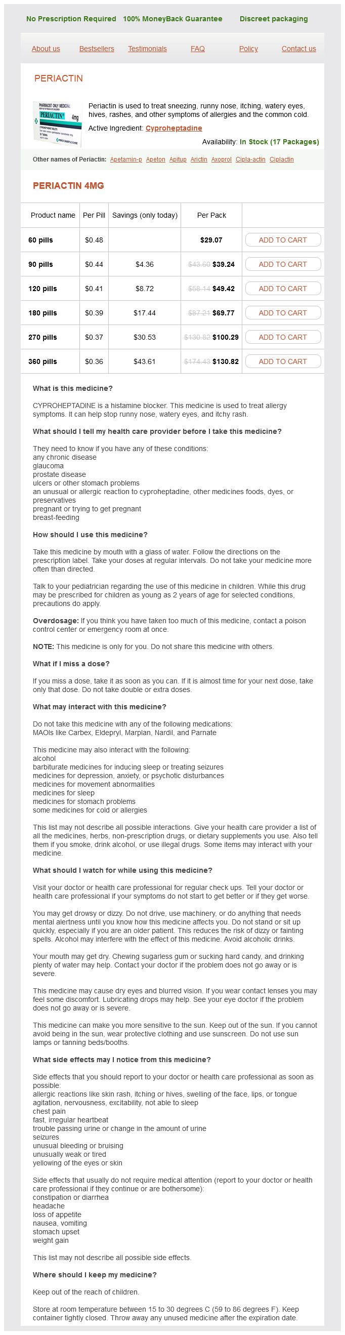
Periactin 4mg
- 60 pills - $29.07
- 90 pills - $39.24
- 120 pills - $49.42
- 180 pills - $69.77
- 270 pills - $100.29
- 360 pills - $130.82
Periactin dosages: 4 mg
Periactin packs: 60 pills, 90 pills, 120 pills, 180 pills, 270 pills, 360 pills
In stock: 752
Only $0.39 per item
Description
Malignant Tumors Kaposi sarcoma Kaposi sarcoma allergy testing greenville nc purchase periactin 4 mg without a prescription, a malignant neoplasm of vascular endothelium, involves the skin and mucous membranes. In the conjunctiva, Kaposi sarcoma presents as a reddish, highly vascular subconjunctival lesion that may simulate a subconjunctival hemorrhage. Options for controlling symptoms include surgical debulking, cryotherapy, and radiotherapy. Lymphatic and Lymphocytic Tumors Lymphoid tumors of the conjunctiva may be benign, malignant, or indeterminate. Lymphangiectasia and Lymphangioma Lymphangiectasia appears in the eye as irregularly dilated lymphatic channels in the bulbar conjunctiva. Anomalous communication with a venule can lead to spontaneous filling of the lymphatic vessels with blood. Lymphangiectasia must be distinguished from ataxia-telangiectasia (Louis-Bar syndrome), in which the epibulbar and interpalpebral telangiectasia of the arteries lacks an associated lymphatic component. The epibulbar vascular lesions of Louis-Bar syndrome can grow with the patient and the eyeball, but episodes of hemorrhage or swelling do not occur. Like a capillary hemangioma, a lymphangioma is usually present at birth and may enlarge slowly. Intralesional hemorrhage, producing a "chocolate cyst," makes differentiation from a hemangioma difficult. Lymphoid Hyperplasia Formerly called reactive hyperplasia, this benign-appearing accumulation of lymphocytes and other leukocytes may represent a low-grade B-cell lymphoma. Most patients are older than 40 years, although, in rare instances, extranodal lymphoid hyperplasia has occurred in children. Biopsy specimens require special handling to complete many of the histochemical and immunologic studies. Fresh tissue is required for immunohistochemistry, flow cytometry, and gene rearrangement studies. Because a patient with an apparently benign polyclonal lymphoid lesion has the potential to develop a systemic lymphoma, general medical consultation is advisable. Lymphoma A neoplastic lymphoid lesion of the conjunctiva is generally a monoclonal proliferation of B lymphocytes. Some lymphomas are limited to the conjunctiva; others occur in conjunction with systemic malignant lymphoma. Some are polyclonal, but most conjunctival lymphomas are monoclonal B-cell lymphomas. Conjunctival plasmacytoma, Hodgkin lymphoma, and T-cell lymphomas are less common. An epibulbar mass fixed to the underlying sclera may be a sign of extrascleral extension of uveal lymphoid neoplasia. Most patients with conjunctival lymphoma are either older than 50 years or immunosuppressed. Unless a tumor is small enough to be removed completely, incisional biopsy is indicated for histologic diagnosis.
Chekan (Cheken). Periactin.
- What is Cheken?
- Cough, high cholesterol, diarrhea, fever, gout, high blood pressure, and other conditions.
- How does Cheken work?
- Dosing considerations for Cheken.
- Are there safety concerns?
Source: http://www.rxlist.com/script/main/art.asp?articlekey=96413
Complications of aqueous paracentesis may include anterior chamber hemorrhage allergy treatment brea ca generic periactin 4 mg with visa, endophthalmitis, and damage to the iris or lens. When the differential diagnosis of the uveitic entity is broader and a larger ocular fluid sample is required, vitreous biopsy should be considered. Vitreous biopsy in selected patients, with carefully planned cytologic, cytofluorographic, and microbiologic examination of vitreous fluid, can be an effective means of confirming a clinical diagnosis. If the results of diagnostic vitrectomy (vitreous biopsy) could potentially alter management of the uveitis, it must be considered. The most common indications include suspected endophthalmitis, primary intraocular lymphoma or other intraocular malignancy, and infectious etiologies of posterior uveitis or panuveitis. In all these scenarios, undiluted vitreous specimens are typically required for testing. Complications of diagnostic vitrectomy in uveitic eyes can include retinal tears or detachment, suprachoroidal or vitreous hemorrhage, and worsening of cataract or inflammation. Although vitreous surgery can be therapeutic and diagnostic in cases of uveitis, the pharmacokinetics of delivered intravitreal drugs are markedly altered in eyes that have undergone pars plana vitrectomy; the half-life of intravitreal corticosteroids, for example, is markedly reduced in vitrectomized eyes. Chorioretinal biopsy, a more technically challenging procedure, may be useful when the diagnosis cannot be confirmed on the basis of clinical appearance or other laboratory investigations. Rapidly progressive posterior uveitic or panuveitic entities, such as a necrotizing retinitis for which the etiology is unknown and the therapeutic regimen undetermined, may require chorioretinal biopsy. Suspected intraocular lymphoma confined to the subretinal space is also an indication for a chorioretinal biopsy. This procedure is performed only after all other less-invasive measures, such as serologic, radiologic, and aqueous and vitreous sample testing, have failed to confirm the diagnosis. It is associated with a high rate of complications and must be performed only by vitreoretinal surgeons with extensive experience using these techniques. Polymerase chain reaction and Goldmann-Witmer coefficient analysis are complementary for the diagnosis of infectious uveitis. Polymerase chain reaction analysis of aqueous and vitreous specimens in the diagnosis of posterior segment infectious uveitis. Diagnostic utility of polymerase chain reaction on intraocular specimens to establish the etiology of infectious endophthalmitis. Therapy Therapy for uveitis ranges from simple observation to complex medical or surgical intervention. Many patients with mild, self-limiting anterior uveitis need no referral to a uveitis specialist. However, in uveitis that is chronic or difficult to treat, early referral to a uveitis specialist may be helpful, not only in eliciting the cause and determining a therapeutic regimen but also in reassuring the patient that all avenues are being explored.
Specifications/Details
The illumination unit is essentially a projector with a light beam that is adjustable in width allergy forecast yonkers ny periactin 4 mg buy visa, height, direction, intensity, and color. The illumination and microscope arms are parfocal, arranged so that both focus on the same spot, with the slit beam centered in the field of view. This setup provides direct illumination, and purposeful shifting of alignment allows for indirect illumination. Direct Illumination Methods Diffuse illumination With diffuse illumination, the light beam is broadened, reduced in intensity, and directed at the eye from an oblique angle. Swinging the illuminator arm to produce highlights and shadows can enhance the visibility of raised lesions of the ocular surface and iris. Focal illumination With focal illumination, the light and the microscope are focused on the same spot, and the slit aperture is adjusted from wide to narrow. Broad-beam illumination, using a slit width of around 3 mm, is optimal to visualize eyelid lesions as well as the corneal opacities seen in dystrophies or scarring. The examiner can use a very narrow slit beam to help identify refractive index differences in transparent structures as light rays pass through the cornea, anterior chamber, and lens. The examiner can also reduce the height of a narrow beam to determine the presence and amount of cell and flare in the anterior chamber. Specular reflection Specular reflections are normal light reflexes bouncing off a surface. An example is the bright round or oval spot seen reflected from the ocular surface in a typical flash photograph of an eye. These mirror images of the light source can be annoying, and it is tempting to ignore them during slit-lamp examination. However, the clarity and sharpness of these reflections from the tear film give clues to the condition of the underlying tissue. Following are the steps for examining the corneal endothelium with specular reflection: 1. Begin by setting the slit-beam arm at an angle of 60° from the viewing arm and using a short slit or 0. Use the joystick to move the biomicroscope slightly forward in order to focus the endothelial reflex. A setting of ×25 to ×40 is usually needed to obtain a clear view of the endothelial mosaic. Cell density and morphology are noted; guttae and keratic precipitates appear as nonreflective dark areas. Indirect Illumination Methods Proximal illumination Turning a knob on the illumination arm slightly decenters the light beam from its isocentric position, causing the light beam and the microscope to be focused at different but adjacent spots. This technique, proximal illumination, highlights an existing opacity against deeper tissue layers and allows the examiner to see small irregularities that have a refractive index similar to that of their surroundings. Moving the light beam back and forth in small oscillations can help the examiner detect small 3-dimensional lesions such as a corneal foreign body.
Syndromes
- Polymyositis
- Antibodies from donated blood samples (intravenous immune globulin)
- Infection (a slight risk any time the skin is broken)
- The opening to the womb (cervix)
- Time it was swallowed
- Shortness of breath
Related Products
Additional information:
Usage: q._h.
Tags: periactin 4 mg purchase visa, cheap periactin 4 mg otc, periactin 4 mg purchase without prescription, generic 4 mg periactin otc
9 of 10
Votes: 132 votes
Total customer reviews: 132
Customer Reviews
Goose, 28 years: Trophozoites and cysts can be observed in greater numbers within areas of retinitis, and T gondii organisms can occasionally be noted invading the choroid, a finding not present in immunocompetent patients. Other interventions available for blind eyes are retrobulbar alcohol injection, retrobulbar chlorpromazine injection, or enucleation.
Hamid, 41 years: Any corneal perforation must first be sealed, or it will continue to leak under the flap. Long-term visual prognosis in children after corneal transplant surgery for Peters anomaly type I.
Gonzales, 52 years: This example emphasizes the importance of the interactions between the central and peripheral chemoreceptors. Furthermore, mechanical debridement may be beneficial for cases of superficial fungal keratitis.
Jarock, 64 years: External ocular and anterior segment findings include scleritis, phlyctenulosis, interstitial keratitis, corneal infiltrates, anterior chamber and iris nodules, and isolated granulomatous anterior uveitis; the last is exceedingly uncommon in the absence of posterior segment disease. Glaucoma Surgery: naturally occurring compound with antibiotic and Principles and Techniques.