- info@careermakers.edu.np
- +977 1 4423870
Doxazosin
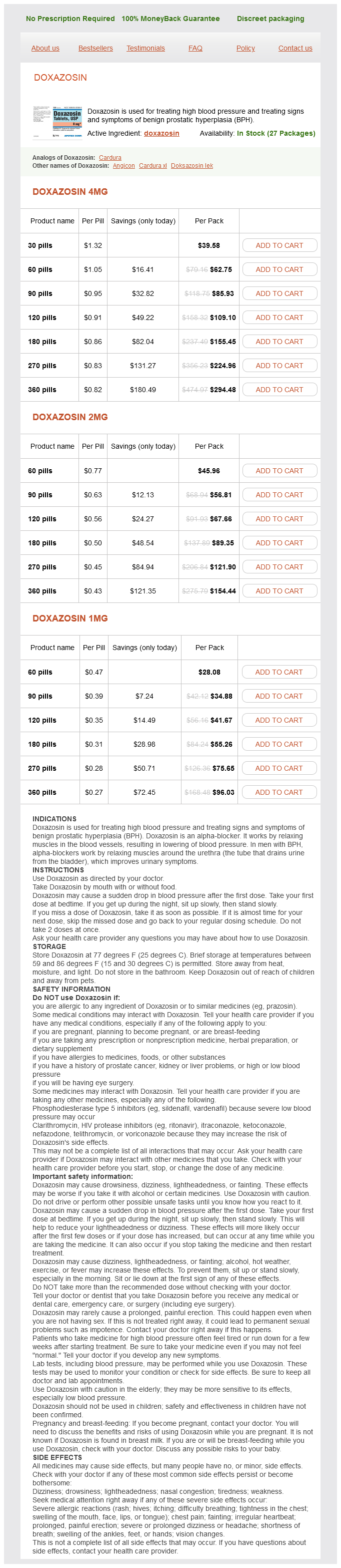
Doxazosin 4mg
- 30 pills - $39.58
- 60 pills - $62.75
- 90 pills - $85.93
- 120 pills - $109.10
- 180 pills - $155.45
- 270 pills - $224.96
- 360 pills - $294.48
Doxazosin 2mg
- 60 pills - $45.96
- 90 pills - $56.81
- 120 pills - $67.66
- 180 pills - $89.35
- 270 pills - $121.90
- 360 pills - $154.44
Doxazosin 1mg
- 60 pills - $28.08
- 90 pills - $34.88
- 120 pills - $41.67
- 180 pills - $55.26
- 270 pills - $75.65
- 360 pills - $96.03
Doxazosin dosages: 4 mg, 2 mg, 1 mg
Doxazosin packs: 30 pills, 60 pills, 90 pills, 120 pills, 180 pills, 270 pills, 360 pills
In stock: 675
Only $0.28 per item
Description
A 1985 report by Ivatury et al compared outcome of patients with a duodenal injury managed by primary repair versus repair with a decompressive enterostomy or serosal patch gastritis zucchini purchase doxazosin 1 mg without prescription, and demonstrates why this type of reconstruction is not recommended. In this report of 60 patients with penetrating duodenal injuries, there was a 64% incidence of abdominal sepsis and a 27% death rate in 11 patients with duodenal gunshot wounds managed with a serosal patch or repair, compared with a 7% abdominal sepsis and 0% mortality rate in 30 patients who had either primary repair or Roux-en-Y anastomotic repair of similar injuries. In 17 patients from that same series with duodenal stab wounds, the complication rate was also higher if the "sucker patch" repair of a duodenal wound was used. The "sucker patch" repair is a modification of the patch repair whereby the open end of a Roux-en-Y loop of small bowel is sutured to the duodenal defect. Given these findings, it is preferable to fully débride the wound, mobilize the edges, and perform a direct end-to-end duodeno-duodeno anastomosis. If adequate mobilization of the duodenal ends is not possible, an end-to-end Roux-en-Y anastomosis should be done. Buttressing the repair with omentum (my preference) or a "serosal patch" from a loop of jejunum seems logical, but the benefit of such techniques is unproved. Diversion of gastric contents is another option, most commonly accomplished by the Vaughan/Jordan pyloric exclusion technique. The concept is to completely divert both gastric and biliary contents away from the duodenal injury, provide enteral nutrition via the gastrojejunostomy, and convert a potential uncontrolled lateral duodenal fistula to a controlled fistula. Although once considered equally effective, the majority of these repairs fail to open, often causing severe complications. The gastrojejunostomy diverts gastric flow away from the duodenum for several weeks while the duodenal and pancreatic injuries heal. The pylorus eventually opens (2 weeks to 2 months) and the gastrojejunostomy functionally closes. In 1993, Asensio and colleagues produced a classical monograph that details all of the surgical techniques in the management of duodenal injuries. Fang and colleagues at Chang-Gung Memorial Hospital in Taiwan have described a technical method of a controlled release of the pyloric exclusion knot and thereby timing the opening of the pyloric occlusion. Marginal ulceration at the site of gastrojejunostomy has been reported in 5% to 33% of patients, prompting some to add truncal vagotomy to the procedure. Most surgeons do not add a truncal vagotomy to pyloric exclusion, however, because nearly all of the pyloric closures open within a few weeks and the occasional marginal ulcer can be medically managed in the interim. The data support the use of pyloric exclusion and gastrojejunostomy in "severe" duodenal injuries or in cases of delayed diagnosis, although no prospective, randomized trial has proved the true benefit of gastric diversion. In addition, the added operating time and the extra anastomosis suggest a good deal of selectivity should be applied to its use. Recent studies have questioned the utility of pyloric exclusion, clearly the most widely used adjunct to duodenal repair. Nassoura and Ivatury and colleagues reported on 66 patients with penetrating duodenal injuries managed between 1986 and 1992; 7 patients died within 48 hours, and of the 59 survivors, 56 patients had primary repair and only 3 (5%) had a pyloric exclusion as an adjunct to combined duodenal and pancreatic injury. These authors recommend use of pyloric exclusion as an adjunct to repair in patients with Penetrating Abdominal Trauma Index greater than 40 or combined pancreatic injury, rather than using the Snyder criteria of Table 1.
Violet Willow (Willow Bark). Doxazosin.
- How does Willow Bark work?
- Treating low back pain.
- What other names is Willow Bark known by?
- Are there safety concerns?
- What is Willow Bark?
- Are there any interactions with medications?
- Osteoarthritis ("wear and tear arthritis"), rheumatoid arthritis, weight loss when taken in combination with other herbs, treating fever, joint pain, and headaches.
- Dosing considerations for Willow Bark.
Source: http://www.rxlist.com/script/main/art.asp?articlekey=96918
It should be noted that obtaining proximal vascular control prior to exploration of a perirenal hematoma is controversial gastritis beer buy doxazosin 4 mg line. Indeed, in one study, preliminary vascular control of the renal hilum had no impact on nephrectomy rate, transfusion requirements, or blood loss. Most surgeons take a more direct approach by simply opening the retroperitoneum lateral to the injured kidney and manually elevating the kidney directly into the wound. A large vascular clamp can be applied proximal to the hilum either at the midline on the left or just lateral to the inferior vena cava on the right to control any further bleeding. Patients who present after blunt trauma may have either a renovascular or renal parenchymal injury, also. Patients in the former group, however, generally present with renovascular occlusion, which will be discussed later. In patients who have suffered blunt abdominal trauma and have undergone imaging that has demonstrated flow to the kidney, there is no justification for exploring the perirenal hematoma should an emergency laparotomy be indicated for other reasons. If a hematoma or hemorrhage is present after penetrating trauma, compression with a laparotomy pad or finger or simply grabbing the bleeding vessels with a hand should be performed as proximal and distal vascular control is attained. The proximal common iliac arteries are exposed by eviscerating the small bowel to the right and dividing the midline retroperitoneum over the aortic bifurcation. In young trauma patients, there is usually no adherence between the common iliac artery and vein in this location, and vessel loops can be passed rapidly around the proximal arteries. Distal vascular control is obtained at the point at which the external iliac artery comes out of the pelvis proximal to the inguinal ligament. Medial rotation of the rightsided abdominal viscera (except the kidney) allows for visualization of the entire infrahepatic inferior vena cava. This artery can be exposed by further opening the retroperitoneum on the side of the pelvis, elevating the vascular tapes on the proximal common iliac and distal external iliac arteries, and clamping or ligating and dividing the large branch of the iliac artery that descends into the pelvis. When bilateral iliac vascular injuries are present, the technique of total pelvic isolation, which includes cross-clamping of the aorta and inferior vena cava just above their bifurcations and distal cross-clamping of the external iliac vessels, will significantly decrease backbleeding from the internal iliac vessels. Injuries to the iliac veins are exposed through a technique similar to that described for injuries to the iliac arteries. It is not usually necessary to pass vessel loops around these vessels, however, because they are readily compressible. As previously noted, the somewhat inaccessible location of the right common iliac vein has led to the suggested temporary transection of the right common iliac artery or ligation of the ipsilateral internal iliac artery in order to improve exposure at this location. Porta Hepatis A vascular injury in the porta may be in combination with an injury to the common bile duct, so some care must be taken with exposures in this area. When a hematoma is present, the proximal hepatoduodenal ligament should be looped with a vascular tape or a noncrushing vascular clamp should be applied (the Pringle maneuver) before the hematoma is entered. If hemorrhage is occurring, finger compression of the bleeding vessels will suffice until the vascular clamp is in place.
Specifications/Details
A ring of bone consisting of two innominate bones and the sacrum forms the pelvis gastritis remedies diet doxazosin 1 mg on line, but there is no 407. Petrone P, Inaba K, Wasserberg N, et al: Perineal injuries at a large urban trauma center: injury patterns and outcome. The innominate bones and the sacrum are held in a structural unit primarily by the ligaments of the pelvis. These ligamentous complexes provide stability for the articulations of the pelvis. Because of the anatomy of the pelvis, some mechanisms are much more likely to produce pelvic fractures than are others. Vehicular crashes are the most common cause of pelvic injury in most environments. They are followed in order of frequency by automobilepedestrian collisions, falls from a height, motorcycle crashes, and crush injuries. Owing to a variety of age-related changes in the pelvis, lower energy mechanisms such as minor motor vehicle crashes and mechanical falls are frequently seen as the source of pelvic fractures in geriatric patients. Although typically not associated with the degree of bony instability seen with high-energy fracture patterns, these fractures may be associated with significant hemorrhage and hemodynamic instability. In the patient presenting with polytrauma, identification of the source of bleeding is of the greatest importance. Up to 20% of patients with pelvic fractures will have concomitant thoracic injuries. The diagnosis of intrathoracic bleeding can be made with a combination of physical examination and a chest radiograph or ultrasound examination. Tension pneumothorax and massive hemothorax can be identified rapidly and treated with tube thoracostomy. Muscle compartment bleeding should be readily identifiable on physical examination, and external blood loss can generally be diagnosed by history from prehospital providers and physical examination. One should keep in mind that external blood loss may become much more apparent once the hypotensive patient is resuscitated and blood pressure increases. The distinction between intra-abdominal and retroperitoneal bleeding can be most difficult. Physical findings such as abdominal pain, distention, or tenderness do not differentiate between intraabdominal and retroperitoneal bleeding. Patients can bleed a large volume of blood into the abdomen and retroperitoneum with minimal physical findings.
Syndromes
- Hallucinations
- Phenacemide
- Hematoma (blood accumulating under the skin)
- Bleeding
- Irritability
- Skin lesion biopsy
- Autologous transplants use your own blood cells or bone marrow.
- Tubes are used to re-route the blood through a special pump called a heart-lung bypass machine. This machine adds oxygen to the blood and keeps the blood warm and moving through the rest of the body while the surgeon is repairing the heart.
- Reflux esophagitis
Related Products
Additional information:
Usage: b.i.d.
Tags: buy doxazosin 4 mg line, doxazosin 4 mg purchase, doxazosin 4 mg order line, order doxazosin 4 mg on line
10 of 10
Votes: 146 votes
Total customer reviews: 146
Customer Reviews
Shakyor, 28 years: These patients are often severely dyspneic, precluding their ability to lay supine on a transport gurney, and finally, their chronic congestive failure may predispose them to easy fluid overload with relatively minimal amounts of intravenous fluid therapy. Unrecognized ureteral injuries usually present in a delayed fashion with persistent ileus, fever/sepsis, abdominal/flank pain, leukocytosis, or fistula. Once in position, the bougie is maintained in position by one clinician as a second clinician advances a lubricated endotracheal tube over the bougie, through the cords, and into position in the trachea under laryngoscopic visualization (whenever possible).
Lukjan, 60 years: Thoracic penetrations need ultrasonic visualization of the pericardial space and esophageal contrast radiography if nonoperative therapy is chosen. If no specific leak is documented, and collaterals are noted to drain into the venous system, medical management has a much higher success rate. Patients with equivocal findings on physical examination or with changing findings needed ancillary diagnostic studies to determine whether any clinically significant injuries exist.
Muntasir, 50 years: A, the clavicle and rib cage form a triangle through which the subclavian vein courses. We strongly feel that if used inappropriately, it will lead to the development of significant cardiac dysrhythmias. It is susceptible to injury, hemorrhage, and air embolism by clamping and retraction and should be treated with respect.
Diego, 47 years: There are a few descriptions of the technical challenges associated with these rare injures, probably because the mortality rate is extraordinarily high. However, compared with internal and external fixation, the poorer outcomes and increased systemic complications associated with skeletal traction with enforced recumbency have resulted in its being used only rarely and temporarily in modern trauma centers. An olecranon osteotomy is frequently necessary in order to provide adequate exposure of the articular surface.
Boss, 61 years: The monitoring is best left to another person who should record vital signs continuously. Furthermore, the medial half of the clavicle may be divided and excised to gain exposure to the proximal axillary and the subclavian artery, especially in bleeding patients. The aim of this chapter is to describe the technique, the indications, the controversies, and the evolution of the process.
Bram, 32 years: Another way to determine proper placement of endotracheal tubes is the syringe aspiration technique. The right coronary may be seen coursing from left to right across the anterior wall of the right ventricle from its origin at the aorta. When the procedure is performed under bronchoscopic control, it is possible to ensure that the needle, guidewire, and the dilators are indeed passing into the trachea, as they are supposed to, and not into a false passage within the soft tissues of the neck.