- info@careermakers.edu.np
- +977 1 4423870
Dulcolax
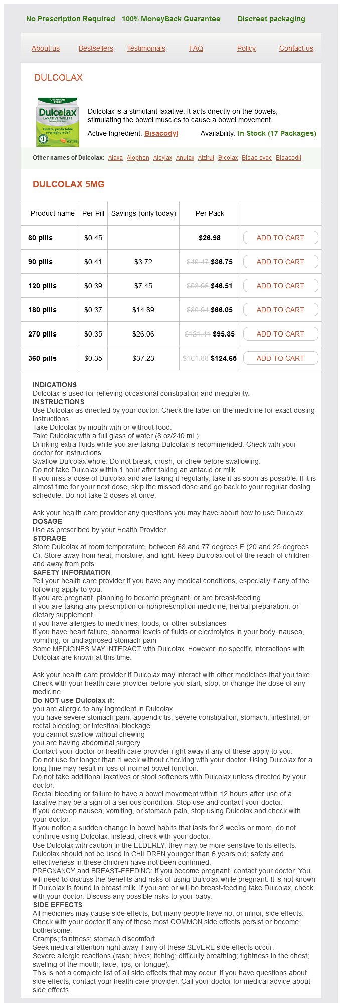
Dulcolax 5mg
- 60 pills - $26.98
- 90 pills - $36.75
- 120 pills - $46.51
- 180 pills - $66.05
- 270 pills - $95.35
- 360 pills - $124.65
Dulcolax dosages: 5 mg
Dulcolax packs: 60 pills, 90 pills, 120 pills, 180 pills, 270 pills, 360 pills
In stock: 758
Only $0.37 per item
Description
These lesions are frequently multiple medicine 014 dulcolax 5 mg order visa, and the differential diagnosis usually includes metastases. When septic emboli induce abscesses, the differentiation from neoplasm becomes much more difficult. It should be noted, however, that other polyphasic disorders such as Lyme disease, sarcoidosis, and vasculitides can appear in a similar manner. The typical differential diagnosis of monophasic white matter lesions includes acute disseminated encephalomyelitis or posttraumatic white matter shearing injuries. Shearing injuries of the white matter almost always have hemorrhage associated with them on susceptibilityweighted images. When hemorrhage is present in white matter lesions, search for a history of trauma. Typically, posttraumatic injuries, migrainous white matter lesions, progressive multifocal leukoencephalopathy, and dilated Virchow-Robin spaces do not enhance. As opposed to demyelinating disorders, the dysmyelinating, peroxisomal, and metabolic disorders are spotted zebras and tend to be bilateral, symmetric, and more diffuse (bizarre). When diffuse white matter disease is associated with macrocephaly,Alexander and Canavan diseases should be suggested. If the white matter abnormality has an occipital predominance and has an enhancing advancing border, adrenal leukodystrophy is the major diagnosis to consider. It is left to serologic and enzymatic analysis to diagnose the most common dysmyelinating disorder, metachromatic leukodystrophy, as well as the lipodystrophies, mucopolysaccharidoses, and other leukodystrophies. Tiny dots of parenchymal hemosiderin staining may also be found with amyloid angiopathy, but you must exclude etiologies such as microbleeds from hypertensive crises, radiation-induced telangiectasias, diffuse axonal injuries, and tiny cavernomas. Use the intraaxial/ extraaxial discussion above to help out (those of you on the electronic version can cut and paste it here). With regard to intradural intramedullary (intraaxial) lesions of the spine, the presence of multiplicity should suggest the diagnosis of a demyelinating disease, sarcoid, hemangioblastomas, neurofibromas, lymphoma, and metastases. Remember to scan the brain for additional lesions depending on the clinical context. Of course, the most common cause of a cord signal abnormality is from spinal stenosis in the cervical spine banging the cord-spondylotic myelopathy. Tumor enhancement is generally focal and nodular rather than diffuse streaky as seen in the demyelinating and inflammatory disorders; however, occasionally, unusual spinal lesions such as herpes zoster, acute disseminated encephalomyelitis, collagen vascular disease, or tuberculosis simulate a neoplasm. Because a dural vascular malformation in the proper clinical setting (Foix-Alajouanine syndrome) may simulate a neoplasm with progressive myelopathy, this may be your only chance to spare the patient a cord biopsy. Be vigilant, as these lesions favor the lower thoracic region, may or may not expand the cord, and generally are centrally located. Also with multisegment high T2 signal in the cord, consider infarct in your differential; the clinical context will be most useful in this regard. Only broad generalizations can be made in trying to determine the histology of an intramedullary glioma, but you might as well take the plunge.
Sweet Chamomile (Roman Chamomile). Dulcolax.
- Are there safety concerns?
- What is Roman Chamomile?
- Dosing considerations for Roman Chamomile.
- Indigestion, nausea, vomiting, painful periods, sore throat, sinusitis, eczema, wounds, sore nipples and gums, liver and gallbladder problems, frostbite, diaper rash, hemorrhoids, and other conditions.
- How does Roman Chamomile work?
Source: http://www.rxlist.com/script/main/art.asp?articlekey=96734
Placenta Previa Previa is the dominant modern risk factor for placenta accreta medications erectile dysfunction purchase dulcolax 5 mg line, with reported odds ratios greater than 50. Prior Cesarean Section A history of one or multiple cesareans has been linked to accreta risk, with some showing a linear increase in both previa and accreta risk based on the number of prior cesarean sections. For example, accreta was found to be more likely when the prior hysterotomy was closed with continuous rather than interrupted sutures32 or when the prior cesarean was elective (unlabored) versus emergent. Nevertheless, the potential to reduce later risk with surgical technique is encouraging. Accreta risk factors beyond previa and prior cesarean section have been demonstrated less consistently. Using a large, multicenter cohort of women undergoing cesarean deliveries, Bowman et al. To date the pathophysiology and clinical behavior of accreta outside of the context of placenta previa is less well defined. Maternal Age Advanced maternal age, defined as 35 years old or greater, is commonly implicated in accreta risk. While it may be a marker for higher parity and placenta previa risk,36 some have found it to be an independent accreta risk factor. This included a range of prior procedures, including myomectomy, uterine curettage, and termination of pregnancy. It also included manual extraction of a placenta and removal of retained products of conception, which could be markers for prior placenta accreta. One study showed a significant trend as the number of curettages increased to three or more. Past Obstetric History Besides history of cesarean section, identifiable risk factors in a past pregnancy may help predict placenta accreta. This variable has received little attention, given that accreta often results in hysterectomy. However, as uterine conservation and accreta without previa is increasingly performed and reported, outcomes in subsequent pregnancies are increasingly described. In 30 women undergoing cesarean section with conservative management of accreta, Eshkoli et al. Placenta Accreta: Epidemiology and Risk Factors 7 Current Pregnancy Factors Given the importance of antepartum accreta identification and delivery planning, factors beyond second and third trimester ultrasound findings have been evaluated as potential predictors of accreta. While such a finding often leads to pregnancy termination, those who continue their pregnancies should be considered very high risk for placenta accreta.
Specifications/Details
Laminar necrosis can lead to mild hyperdensity to the cortical margin of the infarcted tissue treatment 0f gout dulcolax 5 mg order online. Lacunar infarcts have the same intensity characteristics as cystic encephalomalacia albeit on a smaller scale. F, Note marked T1 cortical hyperintensity within infarct on T1-weighted image because of laminar necrosis, not hemorrhage. The frequency is less than 5% and has a male predominance and a mean age of 65 years. The acute clinical findings include the abrupt onset of posteriorly located headaches, severe vertigo, dysarthria, nausea and vomiting, nystagmus, ipsilateral dysmetria, and unsteadiness of gait. Cerebellar infarction can be treacherous, with delayed alteration of consciousness seen in 90% of patients with mass effect. The cerebellum swells with (1) an infarction involving more than one third of its volume, (2) a basilar artery occlusion with poor collateral supply, (3) an embolus with reperfusion, and (4) a massive superior cerebellar artery infarction. On the other hand, dilatation of the great horizontal fissure and adjacent sulci may produce wedge shaped areas of hypodensity that can be confused with infarction. Lesion is more hypodense than acute infarct and it has irregular, somewhat concave margins. Hydrocephalus as manifested by enlargement of the temporal horns (an early sign of obstructive hydrocephalus) may occur and carries a poor prognosis without decompression or shunting. Subtle imaging characteristics (minimal mass effect and slight enlargement of the ventricles) can rapidly evolve to large volume strokes with compression of the brain stem and cerebellar herniation. The superior vermis can herniate upward through the tentorium, whereas the tonsils and inferior vermis may herniate downward into the foramen magnum. Given that death is a highly probable outcome in these types of acute infarcts, there is a bit more leeway in terms of timing of aggressive recanalization; intervention may be performed even up to 24 hours after symptom onset. Treatment of acute cerebellar infarction producing such mass effect can also involve ventricular drainage and cerebellar/posterior fossa decompression often with bilateral occipital bone craniectomy and/or parenchymal resection. Anoxic injuries occur when there is near complete absence of oxygen in the blood for more than 5 minutes while hypoxia occurs when there is partial but more prolonged hypoxemia. A, Computed tomography scan demonstrates an acute cerebellar infarct having a variegated anterior border, producing significant mass effect with compression of the pons and fourth ventricle. Note complete effacement of the posterior fossa cerebrospinal fluid spaces as well. B, Higher section reveals acute hydrocephalus from compression of the fourth ventricle by the cerebellar mass effect. The superior vermis is also involved (arrows) and the swollen cerebellum compresses the quadrigeminal plate cistern. Anoxia can be seen in cardiac arrest, prolonged seizures, strangulation/hanging, near drowning and smoke/carbon monoxide inhalation.
Syndromes
- Difficulty breathing
- Identify problems of the placenta, uterus, cervix, and ovaries
- Rheumatoid factor and ANA test will be negative.
- Boils, painful, red bumps usually involving a hair follicle
- Belching
- Other symptoms of a ruptured AVM
- Developmental delay
Related Products
Additional information:
Usage: q.i.d.
Tags: buy dulcolax 5 mg on-line, dulcolax 5 mg mastercard, purchase dulcolax 5 mg with visa, discount 5 mg dulcolax
9 of 10
Votes: 56 votes
Total customer reviews: 56
Customer Reviews
Ketil, 44 years: Box 5-3 provides a list of conditions that can produce pachymeningeal versus leptomeningeal enhancement.
Brenton, 33 years: Calcium pyrophosphate deposition disease is another source of thickening of the transverse ligament of the odontoid process (like rheumatoid arthritis), cysts, erosion of the dens, but rarely leads to C1-C2 subluxation.
Gunnar, 23 years: On angiography, these tumors are highly vascular, and are usually supplied by branches of the ascending pharyngeal artery and the internal maxillary artery.
Tufail, 62 years: However, the rate for individual populations can be known when complete series of accreta cases are published.
Temmy, 60 years: B, Axial T2-weighted imaging of the brain demonstrate diffuse thickening of the cortex and shallow sylvian fissures.
Hamid, 29 years: The outflow of the lateral ventricle may be obstructed, leading to trapping of one or both lateral ventricles with noncommunicating hydrocephalus.