- info@careermakers.edu.np
- +977 1 4423870
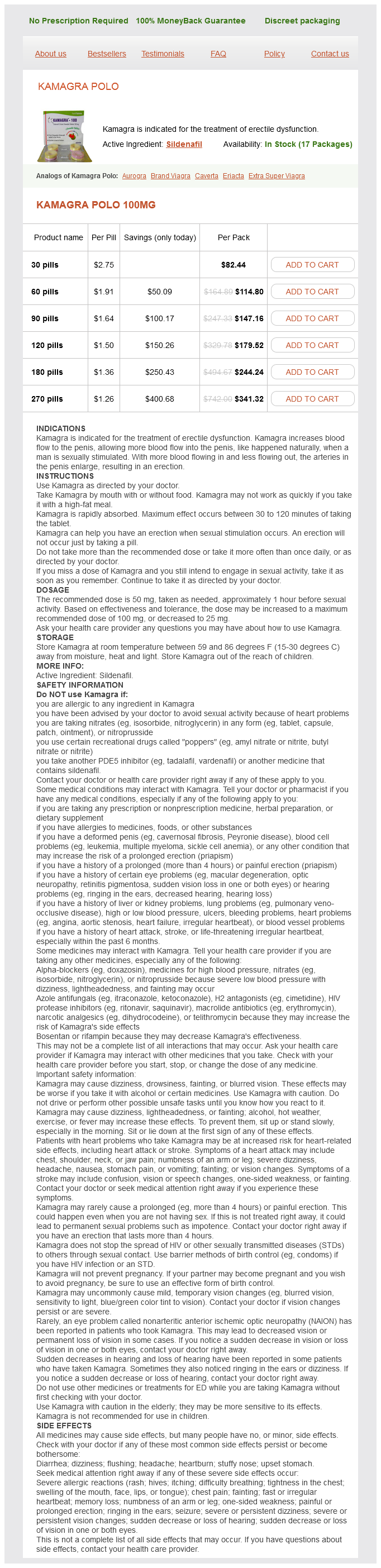
Kamagra Polo
Isaac O. Karikari, MD
- Resident
- Division of Neurosurgery
- Duke University School of Medicine
- Durham, North Carolina
Alcoholism is defined by a combination of criteria erectile dysfunction underlying causes order kamagra polo without a prescription, including the pathological changes just described; physiological tolerance of high concentrations; impaired physiological sublingual erectile dysfunction pills purchase cheap kamagra polo line, psychological erectile dysfunction treatment without medicine generic 100 mg kamagra polo visa, and social functionality; and withdrawal symptoms occurring when intake is reduced or stopped erectile dysfunction pill order kamagra polo 100 mg mastercard. Most alcoholism (type I) sets in after age 25 and is usually associated with stress or peer pressure erectile dysfunction treatment patanjali kamagra polo 100 mg mastercard. These influences lead to increased drinking, which can start a vicious cycle of illness, reduced job performance, family and social problems, arrest, and other stresses leading to still more drinking. Children of alcoholics have a higher than average incidence of becoming alcoholic even when raised by nonalcoholic foster parents. It is by no means inevitable that such people will become alcoholic, but stress or peer pressure can trigger alcoholism more easily in those who are genetically predisposed to it. Alcoholism is treated primarily through behavior modification- abstinence, peer support, avoidance or correction of the stresses that encourage drinking, and sometimes psychotherapy. Drugs such as disulfiram (Antabuse) have been used to support behavior modification programs by producing unpleasant effects from alcohol consumption, but drug treatment has been fraught with potentially dangerous side effects and little evidence of effectiveness. The meaning of dietary fiber; the recommended daily intake of fiber and how actual consumption varies around the world; the forms of dietary fiber; which forms are classified as soluble and insoluble fiber, and differences in the health benefits of these two classes; the detrimental effects of too much dietary fiber 17. Two reasons why the body stores more energy as fat than as carbohydrate; the caloric yield from fat compared to carbohydrate; the typical percentages of body fat in normal males and females 18. Why linoleic acid is called an essential fatty acid; two other fatty acids that might be essential 22. Dietary sources of saturated and unsaturated fats, essential fatty acids, and cholesterol; the health risks of excessive saturated and unsaturated fats 23. Functions of proteins in the body, and a typical percentage of the body mass composed of protein 25. The recommended daily intake of protein; how that can be estimated from body weight; good dietary sources of protein; conditions that call for a protein intake greater than normal; and the risks from excessive dietary protein 26. Why nutritional value of a protein depends on its amino acid composition; the eight essential amino acids; the difference between complete and incomplete proteins; the meaning of net protein utilization and why it is different for dietary proteins of plant and animal origin; and some dietary and ecological advantages of plant protein 27. The meanings of positive and negative nitrogen balance and the conditions under which each of them occurs 28. The definition and ultimate source of dietary minerals; the functions of minerals in the body; the most abundant minerals in the body; and, in general, good and poor dietary sources of minerals 29. The general history of human salt consumption; the recommended daily intake of sodium and some reasons why most U. The definition of vitamins; dietary and nondietary sources; the functions of vitamins in the body; the difference between water-soluble and fat-soluble vitamins Assess Your Learning Outcomes To test your knowledge, discuss the following topics with a study partner or in writing, ideally from memory. Evidence of a homeostatic set point for body weight; relative contributions of heredity and behavior to differences in body weight 2. Sources and actions of leptin and insulin as adiposity signals and long-term regulators of appetite 5. The role of hunger contractions in the onset of feeding; factors that satiate hunger over the short and long terms, and how these resemble the satiation of thirst 7. Hormones that stimulate an appetite for specific classes of nutrients such as carbohydrates, fats, and proteins 8. The definition of calorie and how this relates to dietary Calories (kilocalories) 9. Principal dietary sources of calories; the relative yield from fats as compared to carbohydrates and proteins; and the meaning of empty calories 10. The definition of nutrient; why some nutrients are not digested and yield no calories; and why some things are not considered nutrients even though they are important components of a healthy diet 11. The difference between macronutrients and micronutrients; the nutrients in each category 12. Forms and amounts of stored and mobile carbohydrates in the body; how the body uses carbohydrates; how dietary carbohydrates influence the metabolism of fats 14. Hormones that regulate the balance between blood glucose and stored glycogen; the normal range of blood glucose concentration 15. The recommended daily intake of carbohydrates; the percentage of calories that come from carbohydrates in a typical U. When the body is in its absorptive state; what things occur in this state with respect to carbohydrate, fat, and protein metabolism 2. The main hormone that regulates the absorptive state; its primary metabolic effects in this state; and what antagonist modulates its effects 3. When the body is in its postabsorptive state; what things occur in this state with respect to carbohydrate, fat, and sometimes protein metabolism 4. Hormones that regulate the postabsorptive state, their effects, and the role of the sympathetic nervous system in its regulation 5. How most body heat is produced, and which organs are the most important sources of body heat at rest and in exercise 4. Four mechanisms by which body heat is lost, and the percentages of total heat loss attributable to each of them at rest and at a comfortable ambient temperature (21°C) 5. Two mechanisms for lowering body temperature and two mechanisms of raising it; regions of the hypothalamus involved in each 7. The meanings of nonshivering thermogenesis and behavioral thermoregulation, and examples of the latter 8. Differences between heat cramps, heat exhaustion, and heatstroke; the role of positive feedback loops in hyperthermia; and how and at what temperature range heatstroke can lead to death 9. The role of positive feedback loops in hypothermia, and how and at what temperature range hypothermia can lead to death 26. What cells are primarily responsible for storing and releasing triglycerides; the essence of the lipogenesis and lipolysis carried out by these and other cells 2. The meaning of ketogenesis; the metabolic use of ketone bodies and the pathological effects of excessive ketone levels; common circumstances in which excessive ketogenesis occurs 4. A typical daily rate of protein turnover in the body; where and why the fastest rate of protein turnover occurs; and the dietary and nondietary sources of the amino acids absorbed by the small intestine 5. The meaning of thermoregulation; terms for abnormally low and high body temperatures; and reasons why those two conditions can be fatal Testing Your Recall 1. Which of the following store(s) the greatest amount of energy for the smallest amount of space in the body The brightly colored, iron-containing, electron-transfer molecules of the inner mitochondrial membrane are called 20. The flow of H from the intermembrane space to the mitochondrial matrix creates an electrical current used by the enzyme to make. Extremely low-fat fad diets tend to produce potentially dangerous high levels of blood ketones. Gluconeogenesis occurs especially in the absorptive state during and shortly after a meal. At an air temperature of 21°C (70°F), the body loses more heat as conduction to the surrounding air than by any other means. Also explain whether cyanide poisoning could be treated by giving a patient supplemental oxygen, and justify your answer. Chapter 17 defines and describes some hormone actions that are synergistic and antagonistic. Identify some synergistic and antagonistic hormone interactions in the postabsorptive state of metabolism. Explain why a patient whose liver has been extensively damaged by hepatitis could show elevated concentrations of thyroid hormone and bilirubin in the blood. Your understanding of male reproductive anatomy may benefit if you refresh your memory of the pelvic girdle (see section 8. Sexual development and adult function depend on the gonadotropins and sex steroids introduced in sections 17. F rom all we have learned of the structural and functional complexity of the human body, it seems a wonder that it works at all! The body suffers various degenerative changes as we age, and eventually our time is up and we must say good-bye. In this chapter, we examine some general aspects of human reproductive biology and then focus on the role of the male in reproduction. The next two chapters deal, respectively, with female reproductive function and on the embryonic development of humans and changes at the other end of the life span-the changes of old age. Two things are necessary for reproduction to be successful: (1) gamete motility so they can achieve contact, and (2) enough cytoplasm to divide up into the first cells of a developing embryo, with intracellular nutrients to sustain it until it begins to receive external nutrition. A single cell cannot perform both of these roles optimally, because to contain ample cytoplasm means to be relatively large and heavy, and this is inconsistent with the need for motility. In any sexually reproducing species, by definition, an individual that produces eggs is female and one that produces sperm is male. These criteria are not always that simple, as we see in certain abnormalities in sexual development. In mammals, the female is also the parent that provides a sheltered internal environment for the development and nutrition of the embryo. For fertilization and development to occur in the female, the male must have a copulatory organ, the penis, for introducing his gametes into the female reproductive tract, and the female must have a copulatory organ, the vagina, for receiving the sperm. This is the most obvious difference between the sexes, but appearances can be deceiving (see fig. To achieve this, the parents must produce the reproductive system in the male serves to produce sperm and introduce them into the female body. The female reproductive system produces eggs, receives the sperm, provides a place for the union of these gametes, harbors the fetus, gives birth, and nourishes the offspring. The primary sex organs, or gonads,3 are organs that produce the gametes-testes of the male and ovaries of the female. The secondary sex organs are organs other than gonads that are necessary for reproduction. In the male, they constitute a system of ducts, glands, and the penis, concerned with sperm storage, survival, and delivery. In the female, they include the uterine tubes, uterus, and vagina, concerned with uniting the sperm and egg and harboring the fetus. According to location, the reproductive organs are classified as external and internal genitalia (table 27. Most of them are externally visible, except for the subcutaneous accessory glands of the female perineum. The internal genitalia are located mainly in the pelvic cavity, except for the male testes and some associated ducts contained in the scrotum. The testes produce a normal male level of testosterone, but the target cells lack receptors for it and the testosterone therefore has no effect. In such individuals, the external genitals exhibit female anatomy from birth, as if no testosterone was present. In what way is androgen insensitivity syndrome similar to type 2 diabetes mellitus They typically appear only as an animal approaches sexual maturity (during adolescence in humans). From the call of a bullfrog to the tail of a peacock, these are well known in the animal kingdom. Generally accepted as such in both sexes are the pubic and axillary hair and their associated scent glands, and the pitch of the voice. Other traits commonly regarded as male secondary sex characteristics are the facial hair, relatively coarse and visible hair on the torso and limbs, and the relatively muscular physique. In females, they include the distribution of body fat, enlargement of the breasts (independently of lactation), and relatively hairless appearance of the skin. The distinction begins with the combination of sex chromosomes bequeathed to the zygote. Most of our cells have 23 pairs of chromosomes: 22 pairs of autosomes and 1 pair of sex chromosomes (see fig. Every egg contains an X chromosome, but half of the sperm carry an X and the other half carry a Y. Even an adult male, however, retains a tiny Y-shaped vestige of the paramesonephric ducts, like a vestigial uterus and uterine tubes, in the area of the prostatic urethra. It may seem as if androgens should induce the formation of a male reproductive tract and estrogens induce a female reproductive tract. However, the estrogen level is always high during pregnancy, so if this mechanism were the case, it would feminize all fetuses. Thus, the development of a female results from the absence of androgens, not the presence of estrogens. The sex of a child is determined by whether the egg is fertilized by an X-bearing sperm or a Y-bearing sperm. In the embryo, the genitals begin developing from identical structures in both sexes. It requires an interaction between genes and hormones produced by the mother and fetus. Just as we have seen with other hormones, those involved here require specific receptors on their target cells to exert an effect. Up to a point, a fetus is sexually undifferentiated, or "noncommittal" as to which sex it will become. Its gonads begin to develop at 5 to 6 weeks as gonadal ridges, each lying alongside a primitive kidney, the mesonephros, which later degenerates. In males, the mesonephric ducts develop into the reproductive tract and the paramesonephric ducts degenerate. By 8 to 9 weeks, the male gonadal ridge has become a rudimentary testis that begins to secrete testosterone. Testosterone stimulates the mesonephric duct on its own side to develop into the system of male reproductive posterior to the genital tubercle; and labioscrotal folds, a larger pair of tissue folds lateral to the urogenital folds. By the end of week 9, the fetus begins to show sexual differentiation, and either male or female genitalia are distinctly formed by the end of week 12 (fig. In the female, the three structures just listed become the clitoral glans, labia minora, and labia majora, respectively; all of these are more fully described in the next chapter.

Growth hormone and sex steroids promote protein synthesis and positive nitrogen balance during childhood erectile dysfunction statistics age discount kamagra polo 100 mg overnight delivery, adolescence erectile dysfunction clinics purchase kamagra polo discount, and pregnancy erectile dysfunction specialists cheap kamagra polo 100 mg buy online. Glucocorticoids erectile dysfunction doctor malaysia purchase kamagra polo 100 mg with amex, on the other hand erectile dysfunction underlying causes purchase online kamagra polo, promote protein catabolism and negative nitrogen balance in states of stress. They are required in relatively small quantities, and thus classified as micronutrients. Many mineral salts function as electrolytes and govern the function of nerve and muscle cells, osmotically regulate the content and distribution of water in the body, and maintain blood volume (see chapter 24). Broadly speaking, the best sources of minerals are vegetables, legumes, milk, eggs, fish, shellfish, and some other meats. Cereal grains are a relatively poor source, but processed cereals are often mineral-fortified. Animal tissues contain relatively large amounts of salt, and carnivores rarely lack ample salt in their diets. Plants, however, are relatively poor in salt, so herbivores often must supplement their diet by ingesting salt from the soil. As humans developed agriculture and became more dependent on plants, they also became increasingly dependent on supplemental salt. Salt was once used as a common form of payment for goods and services-the word salary comes from sal (salt). Our fondness for salt and high sensitivity to it undoubtedly stem from its physiological importance and its scarcity in a largely vegetarian diet. Whether salt intake is significantly correlated with hypertension and heart disease has been a subject of great controversy for decades. The kidneys are remarkably capable of excreting excess salt, and multiple studies have found no elevated risk of hypertension in healthy people on high-salt diets. The greatest concern about salt intake arises for patients with renal insufficiency, diabetes, hypertension, and people with a hereditary elevated sensitivity to salt. Hypertension is a leading cause of death among black Americans, for example, who have twice the risk of hypertension and 10 times the risk of dying from it that white Americans have. An evolutionary theory for this is that people of West African ancestry, which includes the majority of black Americans, have kidneys with an especially strong tendency to retain salt. For adults in good health with no hypersensitivity to salt, the Academy of Medicine recommends a daily sodium intake of 1. Liver is specified separately and refers to beef, pork, and chicken livers, which are similar for most nutrients. Vitamin K, pantothenic acid, biotin, and folic acid are produced by the bacteria of the large intestine. Water-soluble vitamins are absorbed with water from the small intestine, dissolve freely in the body fluids, and are quickly excreted by the kidneys. Ascorbic acid promotes hemoglobin synthesis, collagen synthesis, and sound connective tissue structure; it also is an antioxidant that scavenges free radicals and possibly reduces the risk of cancer. The B vitamins function as coenzymes or parts of coenzyme molecules; they assist enzymes by transferring electrons from one metabolic reaction to another, making it possible for enzymes to catalyze these reactions. Some of their functions arise later in this chapter as we consider carbohydrate metabolism. Fat-soluble vitamins are incorporated into lipid micelles in the small intestine and absorbed with dietary lipids. Vitamin A is a component of the visual pigments and promotes proteoglycan synthesis and epithelial maintenance. It is common knowledge that various diseases result from vitamin deficiencies, but it is less commonly known that hypervitaminosis (vitamin excess) also causes disease. A deficiency of vitamin A, for example, can result in night blindness, dry skin and hair, a dry conjunctiva and cloudy cornea, and increased incidence of urinary, digestive, and respiratory infections. An excess of vitamin A, however, may cause anorexia, nausea and vomiting, headache, pain and fragility of the bones, hair loss, an enlarged liver and spleen, and birth defects. Megadoses of fat-soluble vitamins can be especially harmful because they are the most likely to accumulate in the body to toxic levels. Explain the following statement: Cellulose is an important part of a healthy diet but it is not a nutrient. Instead, they transfer the hydrogen atoms to coenzymes, and the coenzymes donate them to other compounds later in one of the reaction pathways. Hydrogen atoms are removed from metabolic intermediates in pairs-that is, two protons and two electrons (2 H+ and 2 e) at a time-and transferred to a coenzyme. This produces a reduced coenzyme with a higher free energy content than it had before the reaction. Coenzymes thus become the temporary carriers of the energy extracted from glucose metabolites. The other proton remains a free hydrogen ion, H+ (or H3O+ when it combines with water, but it is represented in this chapter as H+). Although three monosaccharides are absorbed from digested food-glucose, galactose, and fructose-the last two are quickly converted to glucose, and all oxidative carbohydrate metabolism is essentially a matter of glucose catabolism. Along the pathway of glucose oxidation are several links through which other nutrients-especially fats and amino acids- can also be oxidized as fuel. The numbered steps in this figure correspond to the numbered explanations in the following text. In the body, however, the process is carried out in a series of small steps, each controlled by a separate enzyme. This has two effects: · It keeps the intracellular concentration of glucose low, maintaining a concentration gradient that favors the continued diffusion of more glucose into the cell. In most cells, step 1 is irreversible because the cells lack the enzyme to convert G6P back to glucose. The few exceptions are cells that must be able to release free glucose to the blood: absorptive cells of the small intestine, proximal convoluted tubule cells in the kidney, and liver cells. For now, we are mainly concerned with its further oxidation (glycolysis), the general effect of which is to split G6P (a six-carbon sugar, C6) into two three-carbon (C3) molecules of pyruvate. G6P is rearranged (isomerized) to form fructose 6-phosphate, which is phosphorylated again to form fructose 1,6-diphosphate. This "primes" the process by providing activation energy, somewhat like the heat of a match used to light a fireplace. The "lysis" part of glycolysis occurs when fructose 1,6-diphosphate splits into two three-carbon (C3) molecules. To achieve that in the absence of oxygen, a cell resorts to a one-step reaction called anaerobic fermentation. Most of the matrix reactions constitute a series called the citric acid (Krebs5) cycle. Preceding this, however, are three steps that prepare pyruvate to enter the cycle and thus link glycolysis to it. The acetyl group binds to coenzyme A, a derivative of pantothenic acid (a B vitamin). At this stage, the C2 remnant of the original glucose molecule is ready to enter the citric acid cycle. At the beginning of the citric acid cycle, CoA hands off the acetyl (C2) group to a C4 compound, oxaloacetic acid. Water is removed and the citric acid molecule is reorganized, but still retains its six carbon atoms. No more carbon atoms are removed beyond this point; the substrate remains a series of C4 compounds from here back to the start of the cycle. When oxygen becomes available again, the liver oxidizes lactate back to pyruvate, which can then enter the aerobic pathway described shortly. The liver can also convert lactate back to G6P and can do either of two things with that: (1) Polymerize it to form glycogen for storage, or (2) remove the phosphate group and release free glucose into the blood. Although anaerobic fermentation keeps glycolysis running a little longer, it has some drawbacks. One is that it is wasteful, because most of the energy of glucose is still in the lactate and has contributed no useful work. Skeletal muscle is relatively tolerant of anaerobic fermentation, and cardiac muscle is less so. In the presence of oxygen, pyruvate enters the mitochondria and is oxidized by aerobic respiration. This reaction generates oxaloacetic acid, which is available to start the cycle all over again. The citric acid cycle not only oxidizes glucose metabolites but is also a pathway and source of intermediates for the synthesis of fats and nonessential amino acids. The connections between the citric acid cycle and the metabolism of other nutrients are discussed later. The membrane reactions are carried out by a series of compounds called the mitochondrial electrontransport chain (fig. Ironsulfur (FeS) centers, complexes of iron and sulfur atoms bound to membrane proteins. Unlike the other members, this is a relatively small, mobile molecule that moves about in the membrane. Cytochromes,6 five enzymes with iron cofactors, so named because they are brightly colored in pure form. Hydrogen atoms are split apart as they Cyt a3 transfer from coenzymes to the chain. Transport molecules are reduced when it receives an electron pair and grouped into three enzyme complexes, each of which acts as a proton pump. Molecules at the upper left of the figure have a relatively high free energy content, and molecules oxidized again when it passes the electrons at the lower right are relatively low in energy. Each oxygen atom (half of an O2 molecule) accepts two electrons (2 e) from cytochrome a3 and two protons (2 H+) from the mitochondrial matrix. Without it, this reaction stops and, like a traffic jam, stops all the other processes leading to it. The Chemiosmotic Mechanism Of primary importance is what happens to the energy liberated by the electrons as they pass along the chain. Some of it is unavoidably lost as heat, but some of it drives the respiratory enzyme complexes. Each complex collectively acts as a proton pump that removes H+ from the mitochondrial matrix and pumps it into the space between the inner and outer mitochondrial membranes. Coenzyme Q is a shuttle that transfers electrons from the first pump to the second, and cytochrome c shuttles electrons from the second pump to the third. That is, they create a steep electrochemical gradient across the inner mitochondrial membrane. If the inner membrane were freely permeable to H+, these ions would have a strong tendency to diffuse down this gradient and back into the matrix. As H+ flows through these channels, it creates an electrical current (which, you may recall, is simply moving charged particles). This process is called the chemiosmotic7 mechanism, which suggests the "push" created by the electrochemical H+ gradient. Each enzyme complex pumps hydrogen ions into the space between the mitochondrial membranes. The average adult body contains about 400 to 450 g of glycogen: nearly one-quarter of it in the liver, three-quarters of it in the skeletal muscles, and small amounts in cardiac muscle and other tissues. The enzyme glycogen synthase then cleaves off the phosphate group and attaches the glucose to a growing polysaccharide chain, thus assembling glycogen one glucose at a time. Glycogenolysis, the hydrolysis of glycogen, releases glucose between meals when new glucose is not being ingested. The enzyme glycogen phosphorylase begins by phosphorylating a glucose residue and splitting it off the glycogen molecule as G1P. Liver cells, however, have an enzyme called glucose 6-phosphatase, which removes the phosphate group and produces free glucose. This can diffuse out of the cell into the blood, where it is available to any cells in the body. Gluconeogenesis8 is the synthesis of glucose from noncarbohydrates such as glycerol and amino acids. Explain the origin of the word glycolysis and why this is an appropriate name for the function of that reaction pathway. What important enzyme is found in the inner mitochondrial membrane other than those of the electron-transport chain Describe how the liver responds to (a) an excess and (b) a deficiency of blood glucose. Glucose 6-phosphate Glycogen synthase Glycogenesis Glycogenolysis Glycolysis Glucose 1-phosphate Glycogen Pi phosphorylase Pi Glycogen Key 26. In most cells, the glucose 1-phosphate generated by glycogenolysis can undergo only glycolysis. In liver, kidney, and intestinal cells, it can be converted back to free glucose and released into circulation. These pathways also serve for the oxidation of proteins and lipids as fuel and as a source of metabolic intermediates that can be used for protein and lipid synthesis. Although the total amount of stored fat remains quite constant, there is a continual turnover as lipids are released, transported in the blood, and either oxidized for energy or redeposited in other adipocytes. Glycogenolysis this process and eventually produce just as much glucose as the liver does. Lipogenesis employs compounds such as sugars and amino acids to synthesize glycerol and fatty acids, the triglyceride precursors. As glucose and amino acids enter the citric acid cycle by way of acetyl-CoA, the acetyl-CoA can also be diverted to make fatty acids. The glycerol and fatty acids can then be condensed to form a triglyceride, which can be stored in the adipose tissue or converted to other lipids. When carbohydrate is unavailable, oxaloacetic acid is converted to glucose and becomes unavailable to the citric acid cycle. Fat oxidation then produces excess ketones, leading to elevated blood ketones (ketosis) and potentially to a resulting pH imbalance (ketoacidosis). Name the acidbase imbalance that results from the accumulation of the ketone bodies shown in the oval.
The illuminated spot is then scanned horizontally erectile dysfunction mayo cheap kamagra polo online amex, producing black-and-white images from the epidermis to the upper reticular dermis with an imaging depth of up to 250300 µm erectile dysfunction doctors in lafayette la kamagra polo 100 mg with amex. To visualize a larger area erectile dysfunction treatment jaipur purchase generic kamagra polo on-line, a two-dimensional sequence of images is captured and stitched in a software program to create a mosaic that displays up to 8 × 8 mm of tissue erectile dysfunction doctors phoenix purchase cheap kamagra polo line. In vitiligo lesions erectile dysfunction treatment in vijayawada kamagra polo 100 mg purchase, normal dermal papillary rings disappeared and no bright keratinocytes could be detected in the epidermis [9]. It will be necessary to establish an ideal, meaningful, and reproducible method that is objective, noninvasive, easy to use, and able to assess both morphometric and colorimetric changes during vitiligo treatment in the clinic and in clinical trials. Part of the emitted fluorescence is directed toward the surface of the skin, but it is attenuated by hemoglobin in the capillaries and by epidermal melanin. Woods lamp enhances the contrast between skin with little or no pigment and skin with excessive quantities of melanin. This technique is based on different approaches to the analysis of light reflected by the skin [12]. When light impinges on the skin, approximately 4%8% of the incident radiation is reflected off of the surface. Most of the incident light is attenuated by skin chromophores; that is, melanin, oxyhemoglobin, and deoxyhemoglobin. Oxyhemoglobin and deoxyhemoglobin absorb light specifically between 540 and 575 nm. Melanin heavily absorbs all wavelengths but demonstrates a monotonic increase toward shorter wavelengths [13]. The quality of pigmentation upon treatment cannot be assessed with clinical scoring systems. Demonstration of residual perifollicular pigmentation in localized vitiligo-a reverse and novel application of digital epiluminescence dermoscopy. Application of polarized light dermoscopy in the early diagnosis of vitiligo and its differential diagnosis from other depigmented diseases. Three cases of reverse pigment network on dermatoscopy with three distinctive histopathologic diagnoses. In vivo spectrophotometric evaluation of neoplastic and non-neoplastic skin pigmented lesions. With Fontana-Masson staining, 16% of cases of vitiligo showed the presence of melanin. Skin biopsy samples can also be examined by Melan-A (A103 clone)+ for melanocyte expression by immunohistochemical analysis and for melanin by histochemical studies with section staining by Fontana-Masson method. Melan-A+ cells and melanin granules were detected in depigmented skin as indication that the residual melanocytes are preserved in vitiligo lesions. More than threefold decrease of Melan-A+ melanocytes amount was revealed in perilesional normally pigmented skin of vitiligo patients [3]. Other associated autoimmune diseases include pernicious anemia, Addison disease, and alopecia areata. Patients should be made aware of signs and symptoms that suggest the onset of hypothyroidism, diabetes, or other autoimmune diseases and if any signs or symptoms occur, appropriate tests should be performed. To rule out thyroid dysfunction, thyrotropin testing is the most cost-effective screening test. Clinicians should also consider investigating for serum antithyroglobulin and antithyroid peroxidase antibodies, particularly, as antithyroid peroxidase antibodies are regarded as a sensitive and specific marker of autoimmune thyroid disease. Screening for diabetes can be accomplished with fasting blood glucose or glycosylated hemoglobin testing. Special staining procedures like Fontana-Masson staining is helpful in reaching a definitive diagnosis. Microscopic examination of involved skin shows a complete absence of melanocytes in association with a total loss of epidermal pigmentation. Superficial perivascular and perifollicular lymphocytic infiltrates may be observed at the margins of vitiliginous lesions, consistent with a cell-mediated process destroying melanocytes. Degenerative changes have been documented in keratinocytes and melanocytes in both border lesions and adjacent skin. Other documented changes include increased numbers of Langerhans cells, epidermal vacuolization, and thickening of the basement membrane. Loss of pigment and melanocytes in the epidermis is highlighted by Fontana-Masson staining and immunohistochemistry testing. Patient skin was sampled via a modified suctionblister technique, allowing for minimally invasive, objective assessment of cytokines and T-cell infiltrates in the interstitial skin fluid. There was no significant difference in homocysteine levels between males and females and between patients with negative or positive family histories of vitiligo. Antithyroid peroxidase antibody and antiparietal gastric cell antibody could be useful laboratory markers in a subpopulation of vitiligo patients. However, testing antinuclear antibody, antithyroglobulin antibody, folic acid, and vitamin B12 seems to have limited clinical implications and diagnostic relevance in clinical practice. Both the aforementioned tests could be useful for the characterization of specific subpopulations of vitiligo patients in terms of severity and comorbidity. Lipid disturbances in vitiligo may result from disturbed metabolic processes in the adipose tissue as well as from oxidative stress [7]. Immunohistochemical analysis of melanocyte content in different zones of vitiligo lesions using the Melan-A marker. Reduced immunohistochemical expression of adhesion molecules in vitiligo skin biopsies. Elevated homocysteine levels in suction-induced blister fluid of active vitiligo lesions. Pietrzak A, Bartosiska J, Dybiec E, Chodorowska G, Krasowska D, Hercogova J, and Lotti T. Hepato-splenic and lipid profile abnormalities-do they exist in children affected with vitiligo Melatonin concentration in the blood of vitiligo patients with stress in anamnesis. Detection of serum anti-melanocyte antibodies and identification of related antigens in patients with vitiligo. The residual depigmentation is calculated by a percentage as follows: 100% depigmentation, no pigmentation is present; 90%, specks of pigmentation present; 75%, depigmented area exceeds the pigmented area; 50%, depigmented area equals the pigmented area; 25%, the pigmented area exceeds the depigmented area; 10%, only specks of depigmentation are seen. Also, the course of the disease is varied and includes flareups, remissions, and spontaneous repigmentation. Therefore documentation of the current extent of the disease and also the repigmentation on treatment is extremely important. Various scoring systems are available to assess extent, stability, and repigmentation in vitiligo. In routine practice, the common methods used include visual and photographic analysis. However, these methods are subjective and thus cannot be used in clinical studies. Point counting, digital planimetry, and digital photography with computerized planimetry and computer-aided design software are some of the methods used to assess the area of involvement in vitiligo. Other methods include colorimetry-based image analysis, spectrophotometry, and more modern methods like reflectance confocal microscopy [1,2]. The Wallace rule of nine is a common measure used to measure the extent of involvement in vitiligo [3]. However, assessment of vitiligo severity based on the area of involvement alone is an incomplete measure. The severity of a lesion is much greater when it is located on the face or sexually important sites, such as the genitalia and the breast. This tool has also been widely used to assess the disease response with other modes of treatment. The hands, upper extremities (including axilla), trunk, lower extremities (including buttocks), and feet. For assessment, the body is divided into five units: the head and neck, arms, trunk, legs, and hands and feet. For calculation of extent, the hands and feet are included with arms and legs, respectively. Staging is assessed by cutaneous and hair depigmentation in the largest macule from 04. The staging is done as follows: stage 0: normal pigmentation (no depigmentation); stage 1: incomplete depigmentation (spotty depigmentation, trichrome pigmentation, and lighter homogenous pigmentation); stage 2: complete depigmentation (including hair whitening in <30%); stage 3: complete depigmentation (hair whitening in >30%). The progression/spreading of the lesions is assessed using a simple scale (+1 progressive, 0 = stable, -1: regressive). The limits of the macule are assessed using normal light followed by Woods lamp examination. They are easy to use, can be used in widespread vitiligo, and have been used in clinical studies to assess the response to treatment. Presence of punctate depigmentation, differing shades of hypopigmentation, sharpness of the lesional margins, and Woods lamp examination are some of the criteria used to determine disease stability. The cessation of appearance of new lesions is also taken as criteria for disease stability. Recommendation for vitiligo surgery after 1 year of inactivity is based on this premise. The scoring is as follows: score 4+ activity lasting 6 weeks or less; score 3+ activity lasting 6 weeks to 3 months; score 2 activity lasting 36 months; score 1 activity lasting 612 months; score 0 stable for 1 year or more; score -1 stable with repigmentation for 1 year or more. This measure has also been used for other diseases such as port wine stain and head and neck cancer surgery. The use of individual scales for different parameters is cumbersome in the clinical practice. However, in vitiligo, the emphasis should not only be on measuring the extent of depigmentation; other factors such as quality of life and repigmentation potential also should be considered to gauge a comprehensive picture of the disease. It was developed by Benzekri and coworkers [7] for prediction of repigmentation potential in nonsegmental vitiligo. Each lesion exceeding 10 cm2 is classified based on the clinical type of vitiligo and the melanocyte reservoir (follicular and epidermal) into four types (A, B, C, and D). Comparison of computer-aided design and rule of nines methods in the evaluation of the extent of body involvement in cutaneous lesions. Vitiligo activity index, a new activity evaluation index for bilateral vitiligo vulgaris. Vitiligo Potential Repigmentation Index: A simple clinical score that might predict the ability of vitiligo lesions to repigment under therapy. Autologous transplantation techniques for vitiligo: How to evaluate treatment outcome The assessment measures remain incomplete without the inclusion of this component, as vitiligo has a great impact on the psychosocial aspect of the patient. The disfigurement index measures the alteration in appearance of the patient as perceived by others. It does not cause much physical impairment but causes impairment in mental health and quality of life (QoL) due to its disfiguring appearance, causing discrimination and segregation in certain cultures. Quality-of-life measures evaluate the extent of disability due to a disease in a standardized and quantitative manner. In India, patients with vitiligo face social problems due to its association with social beliefs. Those affected may be unable to find employment, get married, or even get divorced if they develop the disease after marriage. Quality of life is measured in three domains: physical functioning, which includes functional difficulties and symptoms; psychological state, which includes emotional and cognitive functions; and social interaction, which includes public relations, daily activities, and work. Vitiligo affects physical, social, psychological, and occupational aspects of QoL [2]. Due to disgust and fear of infection, these patients are often glared at and avoided. The majority of the studies have found a positive correlation between the body surface area affected by vitiligo and poor QoL, except for few studies by Kent and al-Abadie who found only a weak correlation, and Mishra et al. Have you stayed away from crowded areas (public transportation, shopping centers, etc. Has vitiligo had an impact on what you do on your free time, your activities, and hobbies Have you felt uncomfortable with questions asked about your vitiligo and the explanations you had to make Have you avoided physical contact with others (shake hands, give hugs or kisses, etc. Have you felt uneasy about sharing personal items with the household due to vitiligo Have you had difficulty keeping up with vitiligo therapy (spending too much time or money, applying medicine, etc. Women, patients older than 40 years, and patients with vitiligo located on the feet, legs, and arms were at greater risk for impairment in QoL. Patients with a family history of vitiligo were significantly less worried by their disease in comparison to those with no family history. Friends and family members were perceived as supportive by 80% of the patients, while meeting strangers was uncomfortable as they felt that strangers were less understanding. Patients felt stigmatized by vitiligo, as they often felt stared at, discriminated against, and subjected to rude remarks. They experienced psychological problems such as depression, shame, and anxiety which led to social isolation and low self-esteem. Patients younger than 40 years and those belonging to the working class felt more embarrassed, self-conscious, and discriminated against. However, another study reported that women felt more embarrassed and more self-conscious of their disease, which impaired their social life, sexual activity, personal relationships, and choice of clothing [17]. These patients face restricted career choices, as lesions present on the exposed body sites adversely affect their chances in job interviews. Parents of children with vitiligo have to take leave from work to accompany the child to various appointments [18]. Vitiligo patients had higher prevalence of anxiety or depression (39%) and alexithymia (24%) than the general population. Mood disturbances like irritability and depression were seen especially in teenagers. Children with vitiligo avoided or restricted their sport activities and lost vital days at school.
Discount kamagra polo 100 mg buy. Report Destroys Bill O'Reilly Muslim Hearings Defense.

Syndromes
- Their body language may be unusual.
- Fever
- The major artery from the heart (the aorta)
- Lethargy
- LH
- Get regular exercise, such as walking and pelvic rocking exercises.
- Depressed or angry facial expression
In addition impotence caused by anxiety purchase kamagra polo 100 mg without prescription, some authors have hypothesized and demonstrated that results obtained can be significantly influenced by the blood sample site chosen since diffusion of thyroid hormones and Tg from the thyroid gland may occur at cardiac levels postmortem erectile dysfunction protocol jason discount kamagra polo 100 mg without prescription. Yet again impotence libido kamagra polo 100 mg purchase on line, some authors have recommended the use of femoral vein blood samples concerning postmortem 146 Asphyxiation erectile dysfunction pills side effects buy cheap kamagra polo 100 mg on-line, Suffocation erectile dysfunction medication risks order cheapest kamagra polo and kamagra polo, and Neck Pressure Deaths 19. Thyroid storm induced by trauma due to spear fishing-gun trident impaction in the neck. Postmortem measurements of thyroid hormones in blood and vitreous humor combined with histology. Unreliability of the use of thyroglobulin concentration in postmortem blood samples in forensic diagnosis. The C cells (parafollicular cells) of the thyroid gland and medullary thyroid carcinoma. Lethal pulmonary thromboembolism associated with decreased thyroid hormone levels. Evaluation of postmortem serum and cerebrospinal fluid levels of thyroid-stimulating hormone with special regard to fatal hypothermia. Detection of thyroglobulin in blood stains as an aid in the diagnosis of mechanical asphyxia. Plasma thyroglobulin as an indicator of mechanical asphyxia-comparison of plasma thyroglobulin levels by radioimmunoassay and the results of precipitation-electrophoresis. Concentration of thyroglobulin, thyroxine and triiodothyronine in 3 different causes of death. Circulating thyroid hormone changes in acute trauma: prognostic implications for clinical outcome. Transport of thyroid hormones via the choroid plexus into the brain: the roles of transthyretin and thyroid hormone transmembrane transporters. The analysis of hormones and enzymes extracted from endocrine glands of the neck region in deaths due to hanging. How the reference values for serum parathyroid hormone concentration are (or should be) established Enzyme-linked immunoabsorbent assay for plasma thyroglobulin following compression of the neck. Enzyme-linked immunosorbent assay for determination of plasma thyroglobulin and its application to post-mortem diagnosis of mechanical asphyxia. Thyroid test abnormalities in traumatic brain injury: Correlation with neurologic impairment and sympathetic nervous system activation. Demonstration of thyroglobulin in the heart blood in fatal cases of strangulation. Demonstration of thyroglobulin in the heart blood in fatal cases of strangulation and cut-throat. This means that the toxicology will often have to be taken into account when different alternatives for the cause(s) and manner of death are considered. Since many forms of asphyxia are not incompatible with survival but facilitated by various factors, it is important for the forensic pathologist to review all findings before making the diagnosis. Whereas the toxicological results in complete hangings, or in cases with severe chest compression with unambiguous findings and circumstances, most often do not make any difference to the investigator, the alcohol and drug levels will have to be carefully considered in most other cases of fatal asphyxia. Toxicology in accidental asphyxiations Bolus deaths are regularly encountered by forensic pathologists. The victims are usually either chronic alcoholics or elderly with dementia, or patients with swallowing problems due to neurological disease. In the case of alcoholics, the typical finding is a large piece of food, voraciously swallowed under the influence of alcohol, and sometimes the blood alcohol level is very high. In a study of 78 non-hospitalized persons choking on a bolus of food, one-third were under the influence of alcohol [25]. Although the mechanism underlying bolus death is still not completely elucidated, a vasovagal reflex with cardioinhibitory signalling is one of the most likely alternatives. The normal counterbalance of this overactivation of the parasympathetic system may then fail at significant blood alcohol levels. Due to a reduced muscle strength, subjects severely inebriated by alcohol will also have problems in efficient coughing. Several of the drugs mentioned below as possible facilitating factors in positional asphyxia may also be considered as contributing to a fatal outcome in bolus emergencies. Aspiration, particularly of gastric contents, is a very common finding at autopsy, but its significance for the cause of death is highly variable. Pathologists may overestimate the importance of an impressive aspiration, where the airways actually might not be completely blocked, whereas significant obstruction of the smaller bronchi may go undetected if the examination of the lungs is not performed accurately. Aspiration should be considered as the main or contributory cause of death only when the small bronchi all the way to the finest branches in the lung parenchyma on both sides are blocked by chyme, blood or other material [18]. Even so, agonal aspiration is very common and may in most instances be just an incidental finding. Acute pulmonary oedema and spots with discolouration on the cut surface of the lungs may be signs supporting the notion that the aspiration was vital and significant. In the latter case, microscopic examination may reveal evidence of inflammatory response with accumulation of neutrophils, suggesting that some time has elapsed between the aspiration and death. If the person was under the influence of alcohol and drugs at the time of the aspiration, then the levels might have dropped until death ensued. On the other hand, blood drug levels may increase due to postmortem redistribution if the inhaled vomits contain high amounts of alcohol or drugs. Hence, aspirates containing drugs may represent an important source of artefactually elevated levels of drugs. This implies that the significance of aspiration, and the possibility of postmortem redistribution, have to be taken into account when determining the cause of death [30]. However, in most cases it is very likely that the person would have died from the toxic effect of the drugs even without the final vomiting. Further, postmortem redistribution from aspirated material in the airways is less likely to affect peripheral venous blood, particularly if appropriately collected [48]. The increased risk for drowning when under the influence of alcohol has long been pointed out by authorities in many countries in order to prevent accidental deaths, particularly during the summer months in temperate climate zone countries (see Table 17. Fortyfour per cent of the accidental drowning victims tested positive for alcohol. In Finland, all drowning deaths between 1970 and 2000 were examined with regard to findings and circumstances [20]. The authors also graded the significance of these drugs and concluded that about 16 per cent of the non-boating victims were significantly influenced by the detected psychotropic drugs at the time for the accident. Another interesting observation in that study was that the proportion of alcohol-positive cases showed a gradual decrease from 80 per cent among those aged 5054 years to less than 20 per cent for those aged 8084 years. This panorama probably reflects the increased risk for a fatal outcome in a drowning situation among elderly, regardless of alcohol intake or not. In a recent study from Hungary, 85 per cent of the male drowning victims and 52 per cent of the female victims had consumed alcohol [32]. Lower proportions, of 44 per cent, 41 per cent, 40 per cent and 33 per cent, of alcohol-associated drownings were reported from New York State [4], California [46], Australia [28] and Alaska [42] respectively. These differences might be partly explained by cultural factors and geographical conditions, but the figures are still high enough to justify continued efforts to reduce these untimely deaths. Bathtub drownings constitute an interpretational problem similar to aspiration of gastric contents in intoxication subjects. The difference is, of course, that an aspiration can produce a prolonged process of impaired respiration and result in a delayed death whereas any drowning will go fairly quickly. The problem, however, is that a person who has taken drugs may commit suicide in the bathtub by intentionally inhaling water or may just wait until reaching a state of reduced consciousness when the drowning will be a more or less passive process. The latter may also be true for unintentional drownings among intoxicated persons. Because there is as yet no published compilation of drug levels found in bathtub victims that could be used as a reference, it seems reasonable to use particular caution when evaluating such cases. Positional asphyxia implies that a person has assumed an unnatural position which compromises their respiration. In some cases, the person is trapped in such a position from which it is difficult to escape, but more commonly the victim has sustained injuries during a fall, or is intoxicated by alcohol or drugs, reducing their ability to change position. In general, alcohol intake is associated with increased risk for accidental deaths, including those caused by fatal asphyxia. In experimental settings, even moderate amounts of alcohol have been shown to reduce the tension of the genioglossal muscle complex in awake subjects, increasing the risk that the tongue will fall back and block the pharynx as the person fails to maintain their alertness [19]. These results were confirmed in other studies, in which the number of sleep apnoea events exceeded the normal limit after similar moderate alcohol intake [24,39]. Thus, there is ample evidence to support the notion that alcohol promotes asphyxiation in cases where the victim has adopted an abnormal position compromising the process of respiration. In addition, the general respiratory depressant effect of alcohol, as well as the reduced strength, and in many cases, the effect of alcohol on judgement and coordination, may contribute to a fatal outcome. It is therefore often justified to consider alcohol as a significant contributory factor in positional asphyxia deaths. Pharmaceutical drugs may also be important in positional asphyxia by attenuating the genioglossal tone and/or by affecting other systems important for airway patency. The pedunculopontine tegmental nucleus has been identified as an important control centre for the regulation of upper airway muscle tone. In addition to these electromyogram recordings, they showed that the same treatments to some extent also modulated the breathing pattern. Similar experiments focusing on the role of the retrotrapezoid nucleus and the Kölliker-Fuse nucleus in the brain stem on the respiratory pattern and the tone of the upper airway muscles were performed by Silva et al. In addition, certain drugs, such as the antipsychotic drug clozapine, can also cause hypersalivation [31]. A person who has adopted an abnormal, yet supine position, and is either comatose or immobilized, may aspirate saliva, and if a drug-induced hypersalivation is present, the amounts may be sufficient to compromise the airway flow and contribute to the asphyxia. However, it is a challenge to estimate the respiratory depressant effect only by reviewing the postmortem blood drug concentrations. In order to be considered significant, the drug concentrations should clearly exceed those that can be seen in deaths where the victim obviously was not incapacitated by drugs [10]. Hence, a humble attitude is warranted when assessing the impact on blood drug levels for the fatal outcome in cases of positional asphyxia. A particular form of positional asphyxia is when the body is lying head-down, or even hanging upside down. A number of case reports have described such cases and the circumstances are typically unique to each case. In more recent publications, the pattern is similar; about half of the victims were seemingly influenced by alcohol or drugs at the time of the incident. The most likely mechanisms suggested to lead to a fatal outcome exhaustion of the blood pressure regulation and failure of diaphragm persistence are both likely to be potentiated by alcohol and drug influence. Given the importance of diaphragm exhaustion for the fatal outcome, drugs that reduce the neural signalling to the diaphragm are likely to facilitate the process. Fatal asphyxiation caused by chest compression, often referred to as traumatic asphyxia, will in most cases lead to death fairly rapidly. The petechiae typically appearing on the head, neck and shoulders are not a proof of a long agony but may be produced within minutes. The possible influence of alcohol or drugs will therefore usually not make much difference to the fatal outcome. However, the presence of alcohol or drugs in such deaths may rather be associated with an increased risk for the event as such by contributing to , for example, incautious operation of a heavy machine, or poor driving ability, regarding traumatic asphyxia due to motor vehicle accidents. In many cases, positional asphyxia has also been suggested to have played an important role in these deaths. A large number of violent persons resisting arrest or emergency care are under the influence of alcohol, yet sudden deaths during apprehension and subdual of drunk subjects is rare. In parallel, many reports dealt with positional asphyxia as a competing cause for these in-custody deaths, and a number of experimental studies on human subjects were performed to evaluate the effects of various restraint measures and body positions on respiratory and circulatory parameters [7,8,33]. However, the limitation of these studies was that all volunteers were healthy subjects and were not under the influence of drugs. During the last two decades, a fairly large number of papers have been published reporting on sudden in-custody deaths associated with excited delirium. Virtually all drugs associated with excited delirium are psychostimulants that result in an increase in extracellular dopamine levels in critical brain regions. The main pathophysiological explanation for the clinical symptoms is a dopaminergic overdrive, due to a failure of the affected person to upregulate the dopamine transporter protein [22], although a general depression of the cholinergic system in the brain probably contributes to the delirious state. In the acute phase, death may be caused by cardiac ischaemia due to a tremendous increase in energy demand during extraordinary exertion and sympatico-mimetic overdrive. Hyperthermia is almost always recorded, and may cause death after a while, if unrecognized, or left uncontrolled. The problem is that many of these deaths occur early and unexpectedly during a violent fight, and hence before any medical examination is possible. However, even if this condition has been observed to cause death in the absence of any intervention [36], in most cases restraints, chest compression and abnormal body position are factors that the pathologist needs to consider when evaluating the case. In a retrospective review spanning 20 years, no unintentional deaths due to plastic bag suffocation were identified in the Milan region [9]. On the other hand, plastic bags as a means for committing suicide has been observed for decades, and the method has even been recommended in books on suicide methods and assisted suicide as well as on web pages, possibly affecting the incidence. In most cases, the plastic bag is simply tightened around the neck, but in a proportion of the cases, gas is introduced through a tube into the bag. Most often such arrangements involve a connection to a helium tube [3,44], but there are also reports on the use of nitrogen [21], propane and butane [9] and even ether or chloroform [49]. In the report on chloroform deaths, one subject had chewed a piece of cotton soaked with chloroform, and another applied impregnated surgical masks around the mouth and nose. Toxicological analysis may reveal the presence of these volatiles, but the interpretation is not straightforward, since the concentrations of each of them is difficult to interpret. However, the mere presence of such volatiles in blood or brain tissue samples will in most cases suggest that the person had been inhaling a substantial amount, and even serve as a support for suicide rather than accidental suffocation.
References
- Burge S, Wedzicha JA. COPD exacerbations: definitions and classifications. Eur Respir J 2003; 21: Suppl. 41, 46s-53s. 56.
- Greenberg SM, Eng JA, Ning M, et al. Hemorrhage burden predicts recurrent intracerebral hemorrhage after lobar hemorrhage. Stroke 2004;35:1415-20.
- Salter RB, editor. Continuous Passive Motion. Baltimore: Williams & Wilkins; 1993.
- Philipart M, Engel J, Zimmerman EG. Gelastic cataplexy in Niemann-Pick disease group C and related variants without generalized sphingomyelinase deficiency. Ann Neurol 1983;14:492.
- Berenson JR, Hillner BE, Kyle RA, et al. American Society of Clinical Oncology clinical practice guidelines: the role of bisphosphonates in multiple myeloma. J Clin Oncol 2002;20(17):3719-3736.
- Sola M, Alberro JA, Fraile M, et al. Complete axillary lymph node dissection versus clinical follow-up in breast cancer patients with sentinel node micrometastasis: final results from the multicenter clinical trial AATRM 048/13/2000.