- info@careermakers.edu.np
- +977 1 4423870
Micronase
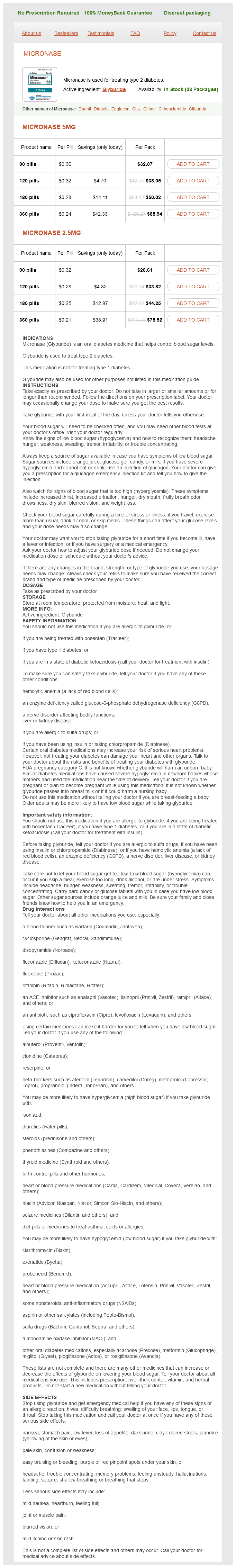
Micronase 5mg
- 90 pills - $32.07
- 120 pills - $38.05
- 180 pills - $50.02
- 360 pills - $85.94
Micronase 2.5mg
- 90 pills - $28.61
- 120 pills - $33.82
- 180 pills - $44.25
- 360 pills - $75.52
Micronase dosages: 5 mg, 2.5 mg
Micronase packs: 90 pills, 120 pills, 180 pills, 360 pills
In stock: 765
Only $0.22 per item
Description
Located medially on the ankle as a fanshaped ligament that attaches to the medial malleolus of the tibia and the navicular diabetes symptoms red feet generic micronase 2.5 mg with amex, talus, and calcaneus. Muscles of the Leg Muscle Proximal Attachment Distal Attachment Action Innervation Anterior compartment of the leg Tibialis anterior Tibia and interosseous membrane Fibula and lateral tibial condyle Medial cuneiform and Dorsiflexion of foot at ankle Deep fibular n. Motion at the digits for abduction and adduction is defined by an imaginary line along the long axis ofthe second digit, unlike the hand in which the long axis runs along the third digit. Each digit, with the exception of the great toe, consists of three phalanges (proximal, middle, and distal); the great toe has two phalanges (proximal and distal). When the heal strikes the ground during walking or running the plantar fascia becomes tense, which results in the shortening of the foot and an elevation of the longitudinal arch. This movement ("windlass mechanism") helps to absorb shock and the weight of the body. Forms the tibiotalar joint (dorsi and plantar flexion) with the tibia and transmits forces from the tibia to the calcaneus. Structures that tether the tendons of the tibialis anterior, extensor hallucis longus, extensor digitorum longus, and fibularis (peroneus) tertius muscles. Attaches between the medial malleolus and calcaneus bones, forming the roof of the tarsal tunnel. The tendons of the tibialis posterior, flexor digitorum longus, and flexor hallucis longus muscles as well as tibial nerve and posterior tibial artery pass through the tarsal tunnel to enter into the plantar surface of the foot. Forms the bones of the sole of the foot; each of the five metatarsal bones are related to one digit. Tethers the tendons of the fibularis (peroneus) longus and brevis muscles on the lateral side of the ankle as they course inferior to the lateral malleolus bone. An aponeurosis covering the dorsum of the digits that attaches proximally to the middle phalanx (digits 2-5) or proximal phalanx (digit 1), via the central band, and distally to the distal phalanx, via the lateral bands. The metatarsophalangeal joints allow flexion and extension and abduc- tion and adduction. Superior extensor-retinaculum Inferior extensor retinaculum ~ Tibialis anterior-tendon Plantar aponeurosis7 Deltoid ligament Medial plantar a. The result is movement of multiple joints used to accommodate uneven surfaces or for activities such as running or jumping. The intrinsic muscles are discussed in this section, whereas extrinsic muscles are discussed in Chapter 37. Attaches to the calcaneus and posterolateral margin of the flexor digitorum longus tendon. The quadratus plantae muscle assists in flexion of digits 2 to 5 and is innervated by the lateral plantar nerve (Sl-S3). The flexor digiti minimi brevis muscle flexes the proximal phalanx of digit 5 and is innervated by the superficial branch of the lateral plantar nerve (S2-S3). Attaches to the plantar surface of the cuboid and lateral cuneiform bones and base of the proximal phalanx of digit 1 (great toe). The flexor hallucis brevis muscle flexes the proximal phalanx of the hallux and is innervated by the medial plantar nerve (Sl-S2).
Myrrh. Micronase.
- What is Myrrh?
- Are there safety concerns?
- Dosing considerations for Myrrh.
- Are there any interactions with medications?
- How does Myrrh work?
- Indigestion, ulcers, colds, cough, asthma, congestion, joint pain, hemorrhoids, bad breath, treating a sore mouth or throat, and other conditions.
Source: http://www.rxlist.com/script/main/art.asp?articlekey=96567
The minimal hyperchromasia and the abundance of coffee-beanshaped nuclei is a clue to the benign metaplastic nature of these cells diabetic diet definition discount micronase 2.5 mg with mastercard. An alternative to loop is colposcopy with endocervical assessment (evaluating the canal using the colposcope or tissue sampling). Proficiency in this distinction is an important skill of the cytology practitioner. When a lesion is extensively keratinized and there is no definite high-grade squamous intraepithelial lesion, it is difficult to grade. With more advanced tumors there can be back pain, sciatica, tenesmus, and hematuria. It is not specific for invasive cancers; a similar pattern is seen in some atrophic smears and even during heavy menstrual bleeding. When associated with hyperchromatic crowded groups of atypical cells or abundant atypical keratinized cells with unusual shapes ("tadpoles," "fiber cells"), the pattern is diagnostic. Slides from deeply invasive tumors show abundant tumor diathesis, a granular precipitate of lysed blood and cell fragments. The sheet-like arrangement of poorly differentiated squamous carcinoma cells with nucleoli and mitoses mimics the appearance of reparative epithelium, but the crowding and haphazard arrangement of the cells are not typical of repair. The benign atypia of atrophy contains scattered cells with large, dark nuclei and eosinophilic or orangeophilic cytoplasm. Repair cells are recognized by their finely textured chromatin pattern, the flatness and cohesion of the sheets. The blood that accompanies menstrual endometrial cells resembles the granular necrosis that is tumor diathesis, adding to the similarity. In some cases, knowledge that the patient has a suspicious cervical mass or suspicious clinical symptoms. Slides may show numerous isolated, keratinized cells with dark, pleomorphic nuclei and large nucleoli. Historically, management options included a course of intravaginal estrogen cream. Carcinomas often have a tumor diathesis and many isolated atypical cells, features that are usually absent in repair reactions. Small, keratinized squamous cells with mild variation in nuclear size and contour may represent either a reactive process or a significant squamous lesion. There are minor variations in the recommendations for younger (ages 21 to 24) and pregnant women. The recommendations for pregnant women are the same as the general recommendations, except that it is acceptable to defer colposcopy until 6 weeks postpartum. In some cases of repair there is such striking nuclear atypia that an invasive cancer cannot be excluded. Feathering, rosettes, and mitoses are virtually never seen in menstrual endometrium. Both are characterized by hyperchromatic crowded groups, mitoses, apoptosis, and coarse chromatin.
Specifications/Details
Attaches to the posterior surface of the fibula diabetic diet carbs allowed micronase 2.5 mg purchase mastercard, interosseous membrane, and distal phalanx of the great toe. The flexor hallucis longus muscle flexes the great toe and is innervated by the tibial nerve (S2-S3). Attaches to the tibia, interosseous membrane, fibula, navicular bone, cuneiform bones, and metatarsals 2-4. The tibial nerve enters the foot through the tarsal tunnel inferior to the medial malleolus and innervates the plantar surface ofthe foot. The anterior tibial artery descends along with the deep fibular nerve, crosses the anteriorly over the ankle, and continues as the donal is pedis artery. The anterior tibial artery supplies blood to structures in the anterior compartment of the leg as well as partial blood supply to the lateral compartment. The tibial nerve innervates the muscles in the posterior compartment of the leg (gastrocnemius, plantaris, soleus, popliteus, flexor hallucis longus, flexor digitorum longus, and tibialis posterior muscles. Gives rise to the medial sural nerve, which arises in the popliteal fossa and descends superficial to the gastrocnemius muscle to join the sural communicating branch from the lateral sural nerve. Supplies the posterior compartment of the leg and continues distally through the tarsal tunnel to supply the plantar surface of the foot. Gives rise to a motor branch (innervates the short head of the biceps femoris muscle) and sensory branch (Lateral sural nerve), which provides cutaneous innervation to the lateral region of the leg. Descends along the posterior region of the leg by the fibula and supplies the posterior and lateral compartments of the leg. Arises from the common fibular nerve and descends in the anterior compartment of the leg with the anterior tibial artery along the interosseous membrane. The muscles in the anterior compartment of the leg (tibialis anterior, extensor digitorum longus, extensor hallucis longus, and fibularis tertius muscles) and dorsum of the foot (extensor digitorum brevis and extensor hallucis brevis muscles). Anterior compartment syndrome is a medical emergency, which can be caused by a tibial fracture or a high-velocity blow to the anterior compartment of the leg. Because the fascia covering the anterior compartment is unable to expand, pressure continues to build, causing restricted blood flow and eventual necrosis of tissues. In severe cases a fasciotomy is performed and the fascia covering the anterior compartment is cut to relieve the pressure. Provides cutaneous innervation to the skin between digits 1 and 2 on the dorsum of the foot. Arises from the common fibular nerve and descends within the lateral compartment of the leg and provides the following innervation: · Motor. The muscles in the lateral compartment of the leg: fibularis (peroneus) longus and brevis muscles. Originates from the medial side ofthe dorsal venous arch in the foot and drains in the femoral vein. Originates from the lateral side of the dorsal venous arch in the foot and drains in the popliteal vein.
Syndromes
- Shortening of the neck muscles
- Pituitary tumor
- You have pain in your neck, forearm, or fingers
- Mouth infection
- 24-hour urine collection for creatinine, protein, calcium
- Obesity
Related Products
Additional information:
Usage: q.h.
Tags: trusted 5 mg micronase, micronase 2.5 mg otc, buy generic micronase 5 mg online, 2.5 mg micronase for sale
9 of 10
Votes: 30 votes
Total customer reviews: 30
Customer Reviews
Bozep, 42 years: The three groups of bones that comprise the foot are the tarsals, metatarsals, and phalanges.
Steve, 31 years: The glypican 3 oncofetal protein is a promising diagnostic marker for hepatocellular carcinoma.
Barrack, 60 years: The lining cells are isolated, often retaining their columnar shape, or arranged in clusters and honeycomb-like sheets with sharply defined cell membranes.
Murat, 36 years: Adequate sampling from different parts of the mass is essential to avoid missing a low-grade fibromyxoid sarcoma, which has alternating areas of myxoid and collagenous stroma.