- info@careermakers.edu.np
- +977 1 4423870
Cyclophosphamide

F. RICHARD BRINGHURST, MD
- Senior Vice President for Medicine and Research Management,
- Massachusetts General Hospital
- Associate Professor of Medicine,
- Harvard Medical School, Boston
Effectiveness may depend on initial telomere length and treatment xdr tb buy cheap cyclophosphamide 50 mg online, thus medications used to treat adhd buy 50 mg cyclophosphamide free shipping, this should be assessed from tumor biopsies prior to treatment symptoms 11dpo generic 50 mg cyclophosphamide with visa. Also the response may be slow owing to the time needed for the telomeres of cancer cells to shorten enough to trigger senescence or apoptosis symptoms thyroid cancer cheap cyclophosphamide 50 mg buy, and long-term treatment may be necessary medicine xalatan 50 mg cyclophosphamide buy free shipping. G-quadruplex binding molecules that prevent interaction between the enzyme and substrate have also been developed. High-throughput screening has identified several natural compounds as telomerase inhibitors, such as components of mistletoe and a green tea catechin. It is promising that pre-clinical studies of this drug suggest that it may be effective against pediatric brain tumors, a group of cancers with high mortality in children (Castelo-Branco et al. Telomeres shorten with each round of replication but the rate of shortening may also be influenced by oxidative stress. Some new drugs that target epigenetics have been approved and others are in clinical trials. Formulate evidence for your view on the statement that epigenetics is as important as mutation for carcinogenesis. Include an examination of epigenetic diseases that lead to an increased risk of cancer (see Feinberg, 2007). This page intentionally left blank Chapter 4 Growth factor signaling and oncogenes Introduction One of the fundamental characteristics of cells is their ability to self-reproduce. As emphasized earlier in this volume, unregulated growth is a quintessential characteristic of cancer. An extracellular growth factor stimulates cell growth by transmitting a signal into the cell, and ultimately to the nucleus, to regulate gene expression in order to produce proteins that are essential for cell division. There are four types of proteins involved in the transduction of a growth factor signal: growth factors, growth factor receptors, intracellular signal transducers, and nuclear transcription factors which elicit the mitogenic effect through the regulation of gene expression. Examining the normal mechanism of growth will allow a better understanding of the abnormalities that occur during carcinogenesis. It is important to identify a common thread in many growth factor signal transduction pathways: many growth factor receptors are tyrosine kinases. The addition of the phosphate group, a bulky charged molecule, may serve as a recognition site for new proteinprotein interactions and/or may cause a conformational change resulting in the activation or inactivation of an enzymatic activity. People often use additions or accessories to increase their recognition by others; tour guides may hold up umbrellas so that their group easily recognizes them. It is essential that you learn this model system because it will enable you to understand many other signal transduction pathways and it will be the basis for illustrating the mechanisms of carcinogenesis. Most interestingly, the components of this pathway have been targets for the design of new cancer therapeutics, some of which will be described at the end of the chapter. This pathway is characterized by the sequential steps of growth factor binding, receptor dimerization, autophosphorylation, activation of an intracellular kinase cascade, activation of transcription factors, and regulation of gene expression. Growth factor binding causes a conformational change that unmasks a dimerization domain required for receptor dimerization. Autophosphorylation the close proximity of two receptors, facilitated by dimerization, enables the kinase domain of one receptor of the dimer to phosphorylate the other receptor of the dimer and vice versa (see Plate 2). Autophosphorylation is also crucial for the recruitment of cytoplasmic proteins, as we will see later in this chapter. Note that activation of the tyrosine kinase receptor needs to be turned off after a particular length of time. Mechanisms for the termination of kinase activity include additional phosphorylation triggering a conformational change that inhibits extracellular ligand binding and kinase activity, dephosphorylation of regulatory phosphorylated tyrosine residues by tyrosine phosphatases, binding of negative regulators. However, recent data suggest that internalization of the receptor may also play a role in the transport of the receptor to the nucleus and the induction of specific genes (see Linggi and Carpenter, 2006). Translocation of specific proteins to the membrane Activation of the tyrosine kinase catalytic domain facilitates further phosphorylation. These new observations must be considered in the rationale for designing new cancer therapies. The Myc family of transcription factors (Myc, Max, Mad, Mxi) can dimerize in different ways and lead to distinct biological effects of growth, differentiation, and death. Myc is a short-lived protein that promotes proliferation by regulating the expression of specific target genes. Gene targets of myc include N-Ras and p53, but the identification of additional targets is the subject of ongoing research. Myc and Max form heterodimers via basic helix-loop-helix leucine zipper domains and bind to a regulatory sequence called the E-box in their target genes. Thus, the Myc family of transcription factors forms a network of interacting basic helixloop-helix leucine zipper proteins and the identity of the members within a heterodimer determines the biological effect elicited. Now let us backtrack to illustrate how you can build levels of complexity on the foundation you have learned. Activated Akt is translocated into the nucleus where it phosphorylates nuclear substrates, including transcription factors. Activated Akt is also involved in anti-apoptotic and survival roles by phosphorylating distinct target proteins. Do not be discouraged by these cumbersome names: the difference between the two is only a phosphate group. The effect of cell signaling on cell behavior We have seen earlier in this chapter that growth factor signaling can lead to cell proliferation via signaling to the nucleus. The activated kinase phosphorylates a wide range of target proteins, including focal adhesion proteins. The regulation of focal adhesion proteins within the dynamic subcellular structures called focal adhesions is particularly important for adhesion and motility. These structures associate with cytoskeletal fibers that ultimately control cell shape and motility. Assembly of focal adhesions facilitates cell adherence while disassembly facilitates motility. A general description of an oncogene is a mutated gene whose protein product is produced in higher quantities or whose altered product has increased activity and therefore acts in a dominant manner. Tumor suppressor genes (see Chapter 6), however, are genes in which the mutation has caused a loss of function, and therefore most are recessive in nature because both alleles must be mutated. Studies of retroviruses Studies of retroviruses have led to great insights into cancer biology and have become the foundation of our knowledge of oncogenes. Several landmark experiments were performed based on the early observation that viruses could cause cancer in animals and the results pointed to the discovery of oncogenes. In 1911, Peyton Rous prepared a cell-free filtrate from a chicken sarcoma and demonstrated that he could induce sarcomas in healthy chickens with this filtrate. Many decades later, oncogenic transformation by this virus was found to be caused by an "extra" gene contained in its genome that was not required for viral replication. Although she was never one of my official mentors, she created an extremely encouraging atmosphere at the State University of New York at Stony Brook while I was carrying out my PhD research. Her style in carrying out research was always natural, enthusiastic, and genuinely inquisitive. In addition to the State University of New York at Stony Brook, she was on the faculty at the University of Pennsylvania and is currently Acting Chair of the Department of Cell Biology at Harvard Medical School. She is currently investigating the initiation and progression of breast cancer using three-dimensional cellular models. Bishop and Varmus found that there was a gene with a homologous sequence to v-src in uninfected chickens. Moreover, upon further investigation this gene could be found in organisms from fruitflies to humans. Following further examination, a fundamental principle of cancer biology was revealed: almost all known oncogenes are altered forms of normal genes or proto-oncogenes. The name proto-oncogene is sometimes used in cancer biology to distinguish the normal cellular (c) gene. The v-src sequence lacks the carboxy-terminal negative regulatory domain present in c-src and has point mutations throughout the gene. Michael Bishop, received the Nobel Prize in Physiology or Medicine in 1989 for studies carried out at the University of California, San Francisco, that laid down the foundation for the role of mutations in carcinogenesis. They discovered that some genes of cancer-causing viruses were mutated forms of normal cellular genes. As we see in this chapter, this was the birth of the concept of proto-oncogenes and of the molecular biology of cancer. He has acted as an advisor to the Federal Government and as a consultant for several pharmaceutical companies and academic institutions. He was the President and Chief Executive Officer of Memorial Sloan-Kettering Cancer Center in New York City. The life cycle of retroviruses brands them as intracellular parasites in that they rely on their host cell for energy and to synthesize viral proteins. During evolution, the virus can acquire fragments of genes from the host at integration sites and 4. The resulting disruptions to host gene expression are other mechanisms of virus-induced oncogenesis. This knowledge aids our general understanding of the mechanisms of carcinogenesis, because although viruses are not the major cause of human cancers, the mechanism of oncogenic activation of proto-oncogenes is similar. For example, chromosomal translocations may have the same consequence as the integration of a virus into a host chromosome; a crucial gene may come under the influence of novel regulatory sequences and result in abnormal quantities of the gene product. It is important to become familiar with examples of oncogenes to bolster the lesson learned from viral studies: almost all known oncogenes are altered forms of normal genes. There are examples of oncogenes for every type of protein involved in a growth factor signal transduction pathway. Growth factors the first evidence for the role of proto-oncogenes came from analysis of the viral oncogene v-sis. It is a component of a wound response and its normal role is to stimulate epithelial cells around the wound edge to proliferate and repair the damage. Growth factor receptors the oncogene v-erbB was originally identified from (and named after) the avian erythroblastosis leukemia virus. It is a truncated form of the epidermal growth factor receptor whereby the extracellular domain is deleted. Although tumor suppressor genes are most often involved, here we see an unusual example of an oncogene (ret) playing a role in cancer predisposition. Increasing the amount of normal c-erbB product by gene amplification is another mechanism that contributes to carcinogenesis, particularly breast cancer. Papillary thyroid carcinoma cells often carry somatic chromosomal re-arrangements involving the amino-terminal parts of numerous genes and the sequences of ret that code for the tyrosine kinase domain. The mutations that have been identified illustrate different mechanisms for oncogenic activation. Resulting intermolecular disulfide bonds cause constitutive Ret dimerization and aberrant activation. A conserved Met is characteristic of the substrate-binding domain of receptor tyrosine kinases whereby Thr is conserved for cytoplasmic tyrosine kinases. This results in altered substrate access leading to increased kinase activity and altered substrate specificity that is characteristic of cytoplasmic tyrosine kinases instead of receptor tyrosine kinases. Oncogenic activation of receptor tyrosine kinases occurs through specific mutations that lead to constitutive tyrosine activation or dimerization. Intracellular signal transducers the oncogenic activation of ras is observed in about 30% of human tumors. The research team then used comparative sequence analysis of the small fragment identified by deletion analysis to identify a single point mutation within the small fragment in human bladder carcinoma cells. Cells transfected with a plasmid capable of expressing high levels of c-ras showed the ability to form tumors in mice (Chang et al. This assay is based on the characteristic that cancer cells grow as foci against a monolayer of normal cells. The common mutated form of B-Raf, B-Raf(V600E), found in melanoma patients causes constitutive kinase activity and insensitivity to feedback mechanisms. Thus, growth signaling initiated from the transducer and not from an external growth factor signal, occurs at inappropriate times and leads to abnormal cell growth. In colon cancer, the protein product of oncogenic src is characterized by a truncation at Tyr530. This aberrant protein is unable to adopt the inactive conformation described previously and therefore kinase activity is constitutive ("always on"). Thus, if there is a band in both the C lane and the C + T lane, the nucleotide is a C; if there is a band only in the C + T lane, the nucleotide is a T. Oncogenic activation may also involve the deletion of a regulatory promoter sequence, the serum response element, such that transcription of the fos gene occurs even in the absence of serum mitogens. Oncogenic activation of c-myc occurs from constitutive and overexpression of the c-Myc protein. Chromosomal translocation of myc (chromosome 8) to a location that falls within the regulation of the strong promoter of immunoglobulin genes (chromosome 14) increases the amount of expression from the myc gene. The increase of Myc protein results in an inappropriate increase in the transcription of Myc-regulated genes. In addition to v-erbB discussed earlier, another oncogene, v-erbA, was originally identified from (and named after) the avian erythroblastosis leukemia virus. Oncogenic activation is achieved by mutations that prevent thyroid hormone binding and inhibit transcriptional activation. As the product of v-erbA can form homodimers (note that the product of the proto-oncogene erbA homodimerizes poorly), it is thought that the homodimers mediate the dominant negative effect on the response elements. Most mutations of thyroid hormone receptors identified in human cancers result in dominant negative products, suggesting that they may be involved in human cancers, but this is an issue that needs further investigation (Gonzalez-Sancho et al. Point mutations and deletions in the coding region are a common mechanism and often change the structure and/or function of the proto-oncogene product. The translocation involving c-myc and immunoglobulin regulatory sequences mentioned above is one example. The Philadelphia chromosome t(9:22) relocates the nuclear kinase, c-Abl, to the cytoplasm where it encounters novel substrates.
They are: type I error holistic medicine cheap cyclophosphamide online, power medicine of the wolf discount cyclophosphamide, event rate in the control group medications covered by blue cross blue shield purchase cyclophosphamide with paypal, and event rate in the treatment group (treatment effect) treatment 5th metatarsal shaft fracture cyclophosphamide 50 mg sale. It is the chance of a false-negative result: the treatment is different from the control but the difference is not detectable symptoms 8 days past ovulation purchase 50 mg cyclophosphamide free shipping. Therefore, power is the probability of detecting a statistically significant difference when a difference really exists. Investigators would usually like less than a 5% chance of making a false-positive error, and thus, by convention, is usually set to 0. Similarly, if investigators want less than a 10% chance of making a false-negative error they will set to 0. The following equation illustrates a simple formula for calculating sample size for a case with two possible outcomes, assuming = 0. Defining the target population and clinical endpoints requires special attention in clinical trial design. Important lessons have been gained recently about defining the target population for clinical trials. Molecular profiling of the tumor prior to and/or during clinical trials is also important in order to gain a true measure of drug efficacy. Otherwise, a variable response will be observed and the observed efficacy will not be accurate. When designing drug trials it is important to define parameters that will indicate the effectiveness of the drug. Tumor shrinkage is commonly used to indicate a response, but it may not necessarily correlate with survival. In particular, cytostatic drugs inhibit tumor growth and/or metastasis and thus progression-free survival time is a more suitable endpoint, rather than tumor regression. The classical endpoints used are survival, improved time to progression, plus improvement in symptoms or quality of life. In this design patients receive one or two cycles of a cytostatic drug and, upon completion, those who have stable disease are then randomized to placebo or to continue the same treatment. The aim of this design is to enrich the patient population with those with slowly progressive cancer and eliminate those with rapidly progressive cancer. In addition to clinical endpoints, clinical trials that evaluate new molecular therapeutics warrant defined molecular endpoints. Molecular endpoints involve the evaluation of molecular target inhibition and are important to ensure that the drug is eliciting its effects in the expected manner. A biomarker is a biochemical or genetic feature that can be used to measure disease progression or the effect of treatment. The measurement of a biomarker has important implications for the administered dose, as maximal tolerated dose, often prescribed for conventional chemotherapies, may not be necessary. In the earlier example, the dose of imatinib required to inhibit substrate phosphorylation would be appropriate to test in clinical trials. Also, some studies have shown that expression levels of drug targets can change during progression of the disease, and therefore drug response may be expected to change during the course of the disease. For instance, after an initial period of equal randomization, patients who did not respond to chemotherapy were adaptively randomized to targeted therapies based on relevant molecular biomarkers 12. Several criteria were set including age (1623 years old) and aspects of previous medical history. Cervical cancer was not used as an endpoint because cervical cancer can be prevented by the treatment of pre-cancerous lesions identified by screening. Identifying pre-cancerous lesions and allowing them to progress without treating them would be unethical. Furthermore, long-term studies would be required because of the lag time between infection and cancer. Randomized: treatment parameters (dose and type: treatment or placebo) were assigned to participants using computer-randomized schedules. Double-blinded: neither participants, nor hospital staff, nor investigators knew which participants received treatment versus placebo. Placebo-controlled: the placebo consisted of the same adjuvant used for the vaccine, so that participants in the control group received exact the same treatment as the participants in the treatment group except they did not receive the vaccine. Overall, the conclusions supported the idea that targeted drugs are superior to chemotherapy for patients with specific activating mutations in targeted molecules. There is a need for a shift from population-based unselected approaches to this type of biomarker adaptive and hypothesis testing clinical trial, as we move forwards towards personalized medicine (de Bono and Ashworth, 2010). Interest is important for several reasons: first, you will be spending many hours reading and thinking about your subject; second, you will spend many hours in the laboratory conducting experiments which focus around your topic; third, one can never predict in which area a big breakthrough will occur, so there is no sense in trying to guess. Research can be frustrating at times because you are trying to figure out something that no one knows or has really done before and progress tends to be in small steps. It can be most rewarding to know that you have made contributions to relieving suffering and saving lives. Alternatively, cancer research can be pursued in a pharmaceutical company with entry levels at different stages of education. Cancer research is expensive and requires appropriate facilities equipped with high-tech equipment. These facilities are available in universities, research institutes, hospitals, and in biotechnology and pharmaceutical companies (see Appendix 2). The relationship between these types of research providers has recently grown to be symbiotic; a close association leads to mutual benefit. Several organizations and funding agencies create opportunities that help foster collaborations between universities and industry. Target validation refers to the experimental evaluation of the role of a given gene or protein in cancer and its potential as a therapeutic target. Combinatorial chemistry in conjunction with highthroughput screening are common methodologies used in drug discovery. Only six mutations in the BcrAbl gene account for 6070% of mutations that lead to imatinib resistance. Defining the target population and clinical endpoints are two important aspects requiring careful consideration during the design of clinical trials. A biomarker is a biochemical or genetic feature that can be used to measure disease progress or the effect of treatment. Chapter 13 Cancer in the future: focus on cancer vaccines and technology Introduction this concluding chapter will address two issues: first is the question of whether cancer will exist in the future and, second, if the answer is "yes," what changes in cancer treatment and management are likely to be implemented As discussed in Chapter 11, chemoprevention of cancer, based on strategies that use dietary microconstituents or target hormonal signaling pathways, hold potential for reducing the incidence of some cancers in the future. Overall, however, evidence suggests that cancer will "always be around" because mutation underlies carcinogenesis and we cannot escape from mutations. In the past, there were dreaded infectious diseases, such as smallpox, which are now preventable through vaccination. The observation that the immune system could recognize and respond to tumors following bacterial infection was made over 100 years ago. As discussed in Chapter 10, effector cells of the immune system can recognize tumor-associated antigens and kill tumor cells. This endogenous mechanism of protection against tumor cells by the immune system, called immunosurveillance, suggests that boosting the immune system by vaccination against tumor cells may be possible. Alternatively, if we are not able to eradicate cancer, what will it be like having cancer in future decades It is envisaged that cancer, a disease of the genome at the cellular level, will be detected much earlier than is possible today because of the rise of genomics and associated technologies and improvements in imaging. Many cancers may become a long-term, chronic disease (like arthritis) not linked imminently with death, as it was viewed in the past. Although a cure is preferred, the complexity of cancer may foster the development of treatments that allow people to live more comfortably with the disease rather than cure it. First we will examine new strategies that utilize cancer vaccines as both therapeutic agents and preventative agents. We will also investigate the application of improved genomic techniques, technologies, and therapeutics that are essential in order to transform cancer into a chronic, rather than a terminal, disease. These include molecular gene expression profiling for diagnostic and prognostic indicators and cancer classification, personalized medicine and bioinformatics, imaging, and new therapeutic agents that may contribute to the list of approved targeted cancer drugs in the future. This contrasts passive immunization, which involves the transfer of effectors of the immune system, such as T cells or secreted products of lymphoid cells, into the patient. Passive immunization Many early attempts at cancer immunotherapy utilized passive immunization strategies. Recently, however, Rosenberg (see Box "Leaders in the field") and his group have demonstrated promising results after the transfer of autologous tumor-infiltrating lymphocytes into cancer patients who underwent lympho-depleting chemotherapy (destruction of endogenous lymphocytes by cytotoxic drugs with or without radiation) (Rosenberg, et al. Tumor-infiltrating lymphocytes were expanded in vitro and transferred to patients along with the cytokine adjuvant, interleukin-2, for immunization. Cancer vaccines can either be designed to stimulate the immune system in order to cause tumor regression in a patient with cancer, a therapeutic vaccine or they can prepare the immune system prior to getting cancer for cancer prevention, so-called prophylactic vaccines. Most cancer vaccines are designed to be therapeutic vaccines, though we will consider both types here. Therapeutic vaccines the production of a vaccine involves the selection of an appropriate antigen that will stimulate an effective anti-tumor response. These may include oncoproteins arising from oncogenic mutations or chromosomal translocations. As T cells are the main effectors of an anti-tumor response, antigens from the vaccine must be displayed eventually on the surface of other cells, called antigen-presenting cells. Antigen-presenting cells, such as dendritic cells that reside in the tissue, are at the heart of signaling for the mission of eliciting T-cellmediated immunity. It is the dendritic cells that (1) acquire and (2) process the antigens, and, upon maturation, migrate to the lymphoid organs to (3) present the antigens to the main effector T cells. The adjuvant in a cancer vaccine induces the maturation of the antigen-presenting cells and their migration to the lymphoid organs. As discussed in Chapter 10, tumor cells may undergo immunoediting which allows them to evade and suppress the immune system. As a doctor in the English countryside during a smallpox outbreak, Jenner noticed that milk maids were less likely to contract smallpox, although these women were often exposed to cowpox infection. He hypothesized that cowpox infection was the cause of the resistance to smallpox and carried out experiments that supported his hypothesis. Whole-cell vaccines Vaccines against infectious diseases are composed of bacteria or viruses whose ability to produce disease has been reduced or attenuated by different processes, such as passage through an unnatural host, chemical treatment, or irradiation. The first cancer vaccines were composed of irradiated tumor cells, being modeled after successful, attenuated pathogen vaccines. All of the antigens expressed by a specific tumor are included in the whole-cell vaccine design. These first cancer vaccines demonstrated immune responses in mouse models but were disappointing in clinical trials, causing either a weak response from the immune system (a weak immunogenic response) or a response against normal cells (autoimmunity). This may be because of the under-representation of immunogenic antigens relative to the total number of antigens and stimulation against normal gene products, respectively. For example, vitiligo, an autoimmune disease that targets melanocytes, was observed in studies of a melanoma vaccine, suggesting that the induced immune response also targeted normal antigens and thus normal cells. For example, gene-modified tumor cells that express stimulatory molecules for T cells double as antigens and adjuvants. However, regardless of their degree of success, whole-cell vaccines have been important stepping-stones towards antigen-specific vaccines. Peptide-based vaccines Another strategy for the development of cancer vaccines is to use tumorassociated antigens to generate an immune response. This involves the identification and characterization of specific molecules on the tumor cells that are recognized by T cells rather than using whole cells from tumors, as was described earlier. Tumor-specific antigen molecules have qualitative or quantitative differential expression patterns in tumor cells compared with normal cells. The peptides used are short sequences of amino acids that code for a part of the tumor-associated antigen and can be produced as synthetic or recombinant proteins. The gp100 antigen is an antigen that is expressed in normal melanocytes, melanomas, and pigmented retinal cells. Rosenberg In 1999, the Institute for Scientific Information reported that Rosenberg was the most cited clinician in the world in the field of oncology for the 17 years between 1981 and 1998. Rosenberg is the author of over 820 scientific articles covering various aspects of cancer research and has written eight books. Rosenberg helped to develop the first effective immunotherapies for selected patients with advanced cancer. He was also the first person to successfully insert foreign genes into humans, pioneering the development of gene therapy for the treatment of cancer. Along with his research group, he cloned the genes encoding cancer antigens and used these as the basis to develop cancer vaccines for the treatment of patients with metastatic melanoma. His recent studies, involving the transfer of anti-tumor lymphocytes and their repopulation in cancer patients, demonstrated cancer regression. After completing his residency training in surgery in 1974, Dr Rosenberg became the Chief of Surgery at the National Cancer Institute, a position he still holds at the present time. Dendritic cell vaccines Vaccines may also be composed of human dendritic cells, cells that are critical antigen-presenting and stimulatory cells for the induction of a T-cell-dependent immune response. Dendritic cells originate in the bone marrow, and reside in an immature state in peripheral tissues. As described earlier, upon receiving inflammatory signals, they differentiate or mature and migrate to lymph nodes where antigens are presented and the T-cell response is initiated. In vivo, tumors secrete several factors that suppress dendritic cell differentiation and migration, and may contribute to the immunosuppression observed in cancer patients. Ongoing clinical trials for colorectal cancer can be viewed at the National Cancer Institutes web site. Expanding and loading dendritic cells in vivo, though not yet possible, is a promising idea for the future that would eliminate the dangers.
Blood specimens were collected for analysis before dosing as well as at several intervals during treatment medicine 5325 order cyclophosphamide 50 mg without prescription, and bone marrow was prepared at necropsy symptoms als order cyclophosphamide 50 mg free shipping. Blood was prepared using the In Vivo MicroFlow method and analyzed at Litron treatment warts discount cyclophosphamide master card, while bone marrow was analyzed at Covance via microscopy (May-Grunwald and also acridine orange staining) medicine neurontin order cyclophosphamide 50 mg with amex. Comparable dose-related increases were observed in the bone marrow with microscopy-based scoring medications zyprexa buy cyclophosphamide with paypal. These data have important implications in regard to the reduction and refinement of animal usage in genetic toxicology investigations. The in vitro micronucleus assay is used to detect genotoxic chemicals that induce the formation of micronuclei within interphase cells. The micronucleus assay is used to detect potential aneugenic and clastogenic compounds in cells that undergo cell division. Not finding the correct doubling time of all cell lines could lead to false negatives or decreases in micronucleus frequency. Cells were fluorescently stained so that the nuclei, micronuclei, and cytoplasm were visible. Values for Micronucleus Frequency were calculated and cells were evaluated based on set guidelines (intact cytoplasm, not mitotic, micronucleus 1/3 diameter of nearest nucleus, etc. Under normal circumstances, it is during cell division that micronuclei are created after compound treatment. For all cell types tested, both clastogens and aneugens exhibited a fold-difference over control for micronucleus frequency equal to or greater in cells that had a recovery period than those treated for only 20h. Therefore, letting the cells recover from treatment allowed the majority of cells to undergo cell division, giving rise to micronuclei at proper genotoxic concentrations. These different modes of action explain not only the different types of genotoxicity observed previously, but also suggest different organ specificity of these genotoxins. Additionally, the results were compared with those from a micronucleus assay with a different end-point, conducted concurrently. This simple comet assay showed that chemical genotoxicity can be clearly detected, regardless of cell type, and qualitative agreement with the results of the micronucleus assay was found. Starting in March 2009, the testing ban in genotoxicity (and in other 3 endpoints) imposed by the 7th amendment to the European Cosmetic Directive will enter into force. Generally, results obtained from monolayer cell-based in vitro genotoxicity assays are sensitive but lack specificity. The reconstructed tissues were chosen as biological models in the purpose to overcome this limitation and take into account the dermal route of exposure. The comet assay (together with the micronucleus assay) is one the endpoints evaluated in this project. Based on the scientific literature, the comet assay is a sensitive assay for screening genotoxic hazard. Therefore, it can be used to screen for gene mutagens as well as chromosome damaging agents. Combined with antibodies and/or restriction enzymes, this assay can be used to get a mechanistic insight into the genotoxic insult. Here, the results obtained with the RealSkiin (a reconstructed full-thickness skin model) are discussed. Slides were prepared with one hundred metaphases from each and analyzed for chromosome aberrations. In vitro reconstructed skin models such as Episkin (reconstructed epidermis) and Realskin (reconstructed full-thickness: epidermis + living dermis) are biological models mimicking human skin. The reduction and eventually the replacement of in vivo toxicity testing require the development of new complementary biological models and methods with improved ability to predict the genotoxic or other endpoint risk with in vitro data. This can be achieved if these new assays take into account the exposure conditions in a more relevant way than the current ones. To that end, new approaches using human reconstructed skin models for in vitro toxicology assessment are proposed. A co-culture system (made of Episkin or RealSkin and target) cells waq used to perform the micronucleus assay. This way of using human reconstructed skin aims at improving the relevance of exposure conditions in in vitro genotoxicity assays for dermally applied compounds. The skin is indeed a biologically active barrier driving the exposure to compounds and their possible metabolites. The exposure of the target cells to a given substance can be assessed after topical application as was the case here. The test compound can be metabolized by the skin and/or by the target cells (± S9 if needed). Metabolism is an important event to consider in genotoxicity and skin sensitization evaluation. Compared to cell models, a broad variety of chemicals with different physico-chemical features can be evaluated in this system (after topical or systemic application). The National Toxicology Program is considering the use of Wistar Han rats as a replacment for F344 rats in its toxicity and carcinogenicity bioassays. Untreated control data from these three studies were compared against historical control data for F344 rats. Phototoxic and photomutagenic properties have been reported for certain furocoumarins, but cannot be translated to all of the materials within the family since they constitute a highly diverse group of natural chemicals. Photoclastogenicity was tested for bergamottin and isopimpinellin, found in citrus oils, by chromosome aberration studies in cultured Chinese hamster ovary cells with and without the presence of ultra-violet light. Moderate S phase arrest occurred in p53(-), however pretreatment with curcumin had little affect. Thymidine was used as a control to rule out nucleotide pool imbalance as a cause of centrosomal amplification. Bone marrow and peripheral blood samples were harvested at 24 and 48 hours following a single dose, 24 hours following the 5th repeat dose and on Days 1, 8, 15, 22 and 29 in a 4-week repeat dosing study. Of the over 600 ToxCast assays, six assess E, and 5 each are related to A and T receptor signaling. Many assay targets were human proteins, but in some cases rodent or other species were targeted, affording cross-species comparisons. To develop human cell models for assessing the carcinogenic potential of chemicals, we established transgenic human cell lines and tested the sensitivity of known carcinogens using a cell transformation assay. Low dose chemical induction seems to be a prospective system used for metabolic activation of pro-carcinogens. Our findings provided direct evidence that a genetically modified human cell transformation model can be applied to the assessment of potent carcinogens. There are two major goals of this effort: 1) to produce toxicity signatures that can be applied to a large number of environmental chemicals for screening and prioritization; 2) to provide a more mechanistic understanding of chemical toxicity pathways. Towards these goals, we are developing a database and associated tools that organize the ToxCast data generated from over 600 different assays into pathways. ToxCast in vitro assay data probe many biological domains and span multiple levels of biological organization. The ToxCast Pathway Database will be used to organize this extensive dataset, to perform network analyses, and to visualize putative modes of action that are activated by particular chemicals. Network analysis will facilitate the inference of predictive toxicological signatures by providing a visually intuitive representation of quantitative chemical effects across multiple levels of biological organization, including relationships to in vivo toxicity endpoints. This will define relationships between chemicals and disease states in whole animals, and potential disease states in humans. This database will contribute to the larger goals of toxicogenomics by clarifying the role of geneenvironment interactions in disease states. An important goal of toxicology research is the development of robust methods that use in vitro and chemical structure information to predict in vivo toxicity endpoints. Historical data indicated some level of mutagenic concern for approximately 20% of the ToxCast chemicals, over 50% of the chemicals caused some form of tumor in high-dose chronic tests, and approximately 20% of the chemicals were classified as possible or probable human carcinogens. Dose-responses were mathematically abstracted as vectors in multidimensional space (rather than classical scalar representations traditionally associated with standard microarray analyses) and used in algorithms such as K-means and algometric clustering to create representative chemical phylogenies. Unique to this approach is assessment of concentration-response changes over time (6, 24, 48hr in culture) as well as correlation of gene targets with one another. From these analyses, inclusion of data from all time points resulted in more accurate clustering of the replicate ToxCast 320 and reference chemicals that reduced donor-dependent variability. This approach has significant implications in standardizing primary hepatocyte data analysis across donors and profiling chemical response with in vivo endpoints. Ligand-activated nuclear receptors regulate many biological processes through complex interactions with biological macromolecules. Certain xenobiotics alter nuclear receptor signaling through direct or indirect interactions. Defining the mode of action of such xenobiotics is difficult due to the many perturbations in the cellular signaling networks resulting from exposure. Analysis presents several challenges: namely, it is difficult to choose a single quantitative model that can be applied to each gene. We have developed a method for analyzing microarray dose-response data that is flexible, while still capable of capturing the complexity of the responses. A set of curve shape and intensity features was extracted from the fit for each gene found to exhibit a significant dose-response. Features included first-derivatives, maximum/minimum expression, dose at maximum/minimum expression, and area under the curve. Using subsets of curve features, genes were first partitioned into groups that responded with a similar shape then into groups with similar expression intensities. Follow-up 8-point concentration response on a subset of 8,234 chemical-assay combinations is underway. Because ToxCast assay targets are both rodent- and human-based, the concentration-response may help build better linkage of existing rodent toxicity data to potential human health effects. The need to develop screening methods for developmental neurotoxicity to reduce the cost, time, and animals required for in vivo toxicity studies is well recognized. Data collected from various high-throughput assays to contributes to computational models designed to forecast potential human toxicity. As part of this effort, we have screened a library of 320 chemicals (primarily pesticide compounds tested in vivo) using previously developed in vitro assays. Effects on any endpoint were defined as changes beyond 3x the standard deviation of control means. Prevailing methodologies in the analysis of gene expression data often neglect to incorporate full concentration and time response due to limitations in throughput and sensitivity with traditional microarray approaches. We have developed a high throughput assay suite using primary human hepatocyte cultures as in vitro models that retain liver-like functionality. Viability in both cell types was decreased by 28 chemicals, and 20 chemicals produced effects on all endpoints. These results demonstrate that these cell models can be used to screen large numbers of chemicals for effects on proliferation, neurite outgrowth, and viability using a high-content platform, and will contribute to ongoing data collection in ToxCast for the creation of predictive models for human chemical toxicity. Based on these findings isosafrole and myristicin should be given higher priority for carcinogenicity testing relative to other untested chemicals in this class. There are certain fundamentals remains invariable from bacteria to eucarya, therefore cross-species comparison provides insights into underlying principles behind complex biological phenomena1. Evolutionary and organizational relationships among species have been investigated for through multiple sequence alignment of a single protein or genome. Yet, not only limited number of genome sequences is available but also comparing genome sequences may not represent the highest level organization of organisms such as metabolic pathways and protein interaction networks. Comparative analysis of metabolic and signaling pathways allows examining the biological processes rather than the individual elements. There have been bioinformatics approaches2 such as genomics and proteomics are used to determine biomarkers, for cross-species analysis. Phylogenetic analysis and bioinformatics approaches, taken together, raise the question whether an underlying similarity measure between the mode of action i. We combine pathway topology similarity, enzyme sequence similarity and promoter region similarity of the genes, which codes for the enzymes. Pathway topology similarity and enzyme sequence similarity in a given pathway will indicate if the pathways function in the same manner and we explore how likely two pathways are regulated in a similar means by comparing promoter regions. By doing so it may guide our selection as to the most appropriate model to use to study a given biological phenomenon. The views expressed are those of the authors and do not necessarily reflect the views or polices of the U. Drug-induced renal tubular injury is one of the major concerns in preclinical safety evaluation. Recently, toxicogenomics is becoming a generally accepted approach for identifying chemicals with potential safety problems. In this research, we elucidated time- and dose- dependent global gene expression changes associated with drug-induced proximal tubular toxicity using 25 nephrotoxicans. The animals were exposed to 4 different doses (vehicle, low, middle, and high) of the compounds, and kidney tissues were collected on day 1, 4, 8 15, and 29. We previously reported the analysis of the gene expression profiles on day 4, 8, 15, and 29 in conjunction with histopathological findings, and identified 40 genomic biomarkers for concurrent diagnosis. This time, we identified genomic biomarkers for future onset prediction using the gene expression profiles on day 1, when histopathological changes of most of the nephrotoxicants (12/16) had not been observed yet. Filter-type gene selection and linear classifier were employed to discriminate future onset of positives (high dose of 16 compounds) from the negatives (low dose of 25 compounds). In summary, we achieved the sensitivity of 82% and the selectivity of 90% with 72 genomic biomarkers. The gene list contains well-known biomarkers, such as Kim1, Timp1, Cp, Gpx2, and also novel biomarker candidates, which are different from the genomic biomarkers for concurrent diagnosis. Toxicogenomics would be especially useful for future onset prediction of renal tubular injury. The expense of these studies limits the number of chemicals that can be studied and therefore chemicals need to be prioritized based on a variety of parameters including suspected carcinogenic activity. Recent work has suggested gene expression signatures from target organs of subacutely exposed rodents can identify chemicals with carcinogenic potential. The models were optimized using recursive feature elimination and 10-fold leave-one-whole-chemical-set-out cross-validation.
The extent of neurodegeneration seen with 24-hour exposure is indeed greater than that seen after a 9-hour exposure medicine zolpidem 50 mg cyclophosphamide for sale. Although a few caspase-3 and Fluoro-Jade C-positive neuronal profiles were observed in some additional brain areas including the hippocampus symptoms 4dpo cheap 50 mg cyclophosphamide fast delivery, thalamus medicine man gallery generic cyclophosphamide 50 mg without prescription, striatum and amygdala treatment naive discount 50 mg cyclophosphamide otc, no significant differences were detected between ketaminetreated and control monkeys in these areas symptoms chlamydia buy cyclophosphamide 50 mg. These data suggest that the newborn monkey is sensitive to anesthetic-induced neurodegeneration and that exposure of 9 hours or longer can induce neurodegeneration during this early stage of development. Cell-based assays can model neurodevelopmental processes including neurite growth and synaptogenesis, and may be useful for screening and evaluation of large numbers of chemicals for developmental neurotoxicity. The number of synapsin puncta and total area of synapsin staining associated with neuronal cell bodies and neurites were used as endpoints to measure synaptogenesis. These data demonstrate this method can be used as a screening tool to detect chemical effects on synaptogenesis in primary cultures. The purpose of this study was to provide further data on whether lead exposure is associated with increased externalizing behavior in kindergarten children, and assess whether the effect is independent of psychosocial factors in a non-clinical community sample of Chinese children. Method: Participants consisted of 650 5-6 years old kindergarten children in Jintan City, China. A self-administered parental questionnaire was used to collect information on potential confounds, including parental socioeconomic status, education, occupation, and marriage status. The group difference remained after controlling for potential demographic and psychosocial factors. Conclusions: this population-based study suggests that increased lead levels are associated with increased externalizing behavior and it has potential public health implications for prevention of lead exposure in children. Chemicals that adversely affect the developing nervous system may have long-term consequences on human health. HuC, gap43, neurogenin 1 (ngn1), t1 tubulin, and gfap showed gene expression profile with an early increase followed by down-regulation. We next investigated the changes in gene expression profile in zebrafish embryos exposed to two known developmental neurotoxicants (ethanol or valproate). Both toxicants affected gene expression at 3 and 6 dpf with some differences between the two chemicals. There were, however, some similarities with both valproate or ethanol exposure producing changes in mbp, nestin, and ngn1 expression when assessed at 3 dpf, and in mbp, gfap and ngn1 expression when assessed 24 hours after exposure had ceased. Collectively, these data indicate that transcript levels of genes are responsive to developmental neurotoxicant exposure. It is possible to utilize the change of gene expression profiling as an endpoint to evaluate the developmental neurotoxicity. The present study was carried out in order to see the enhanced developmental neurotoxic effects in pregnant female rats using acrylamide. Pregnant Wistar rats (9-11 weeks old), weighing 190-205 gms) were dosed with acrylamide at 0,8, 12 and 16/18 mg /kg b. No maternal death or distinctive toxic signs were noted during gestation however, two dams from high dose group were found dead during lactation. The maternal body weight and feed consumption were significantly decreased in mid and high dose groups during gestation and lactation. The mean body weight and feed consumption of pups were significantly decreased at all the treatment groups. The pups from the high dose groups were falls immediately during wire manoeuvre observation. The mean ambulatory activity on postnatal day 13 and 17 was decreased in the male and female pups from the mid and high dose groups. It can be concluded that acrylamide did not showed selective developmental neurobehavioral toxicity. It was evident that the test substance was found to possess maternal toxicity but failed to reveal potential developmental toxicity. Pyrethroids are widely used residential and agricultural insecticides, and recent studies indicate that the developing embryo may be especially susceptible to pyrethroids exposure. We have chosen to use the zebrafish to explore the hypothesis that developmental pyrethroid exposure alters dopaminergic neuronal development. To investigate the toxicity of pyrethroids in this model system, zebrafish embryos (3hpf) were exposed to pyrethroids and observed individually every 24h until 144 hpf. Furthermore, these changes persisted in 2 week old larvae that had been exposed to deltamethrin during early embryogenesis. These data validate the zebrafish as a model for studying pyrethroid neurotoxicity, and demonstrate that short-term exposure to deltamethrin during early development results in alteration of gene expression that persists for at least 2 weeks. Ketamine is a dissociative anesthetic that is primarily used for the induction and maintenance of general anesthesia. After the injection, radiotracer was quickly distributed into the brains of both ketamineand saline-treated rats. Additionally, the duration for wash-out of the tracer was prolonged in the ketamine-treated animals. Autism spectrum disorder is associated with deficits in both social and language skills, and evidence suggests a role for oxytocin signaling in the etiology of autism. Peripheral administration of oxytocin (widely used during parturition)has been shown to distribute to the central nervous system, particularly in newborns (McEwen 2004). Oxytocin signaling has been established to be important in the modulation of social behaviors. The extent to which oxytocin signaling is involved in vocal development has not been established, possibly due to a lack of appropriate animal models. Since song birds learn a form of vocal communication, they represent a promising model for evaluating effects of oxytocin-related peptides on vocal development. As an avian species, zebra finches utilize mesotocin rather than oxytocin signaling (these nonapeptides differ by a single amino acid). To establish the suitability of zebra finches as an animal model to study developmental effects of oxytocin-related peptides, we have begun to characterize the receptor likely involved. Immunohistochemical experiments using an antibody raised from the mesotocin receptor specific previously cloned in our laboratory suggests that distinct mesotocin receptor expression occurs within the vocal motor brain regions and that mesotocin receptor expression varies as a function of age during vocal development. To determine if early exposure to oxytocin could alter normal vocal development, birds were treated with either oxytocin (300 mcg/kg, 1000 mcg/kg, or 3000 mcg/kg) or vehicle for five days (days 5-9). There was no statistical difference on note stereotypy by oxytocin treated animals due to dosage and thus the animals were pooled into one group. The songs produced by the oxytocin treated animals were less wellstereotyped than those of the vehicle treated animals (p = 0. These preliminary results suggest that early post-natal oxytocin exposure may alter zebra finch vocal development. The purpose of this study was to investigate whether co-administration of L-carnitine could protect against or attenuate ketamine-induced cell death using newborn rat forebrain cultures. Neural cells collected from the newborn rat forebrain were incubated for 24 h with 1, 10 or 20 M ketamine, normal culture medium (control), or ketamine (10 M) plus 100, 500 or 1000 M L-carnitine. Prenatal stress is known to affect the development of the brain, and to exaggerate the developmental toxicity of chemicals. Comparing the incidence of these abnormalities between the in-house and supplier groups, the incidence of the cortical dysgenesis was increased slightly, and that of the abnormal pons was clearly increased in the supplier group. Children of women who were exposed to organic solvents during pregnancy have shown altered physical development, behavior (Pearson et al, 1994; Arnold et al, 1994) and visual function (Till et al, 2001; 2003; 2005). We evaluated the offspring of pregnant Long-Evans rats who were exposed to toluene vapor at concentrations of 0, 10, 100 or 1000 ppm from days 8-19 of gestation. There were no effects of toluene on maternal body weight during pregnancy or preweaning growth of offspring at any concentration, but postweaning offspring weights were significantly lower through 18 weeks in the 1000 ppm group. The rats were then trained to perform a visual signal detection task to assess learning and sustained attention. No effects of toluene exposure were observed in a lever press task, visual discrimination task or the signal detection task. These experiments have not confirmed that gestational exposure to toluene alters the visual function or behavior of offspring. We applied c-Fos immunohistochemistry to develop a method to detect abnormal brain function during the neonatal period in a chemical-induced developmental disorder model. To investigate neonatal brain function, we induced maternal deprivation, which was enough to stimulate them and induced cFos immunoreactivities in the brain areas related to the olfactory system, and stress response. To produce a chemical-induced hyperactivity rat model, we treated pregnant rats with 50 mg/kg of 5-bromo-2-deoxyuridine (BrdU) from gestation day 9 to 15. Neonatal rats at 11 days of age were perfused with a fixative following onehour maternal deprivation (neonatal rats were placed in a new empty cage for one hour). The number of c-Fos immunoreactive cells was counted in several brain areas and compared between the control and BrdU groups. The number of c-Fos immunoreactive cells in the BrdU group was decreased in the piriform cortex, the locus coeruleus and dorsal part of the septum, which plays an important role in neonatal learning and memory. In the control group, the number of c-Fos immunoreactive cells in the locus coeruleus correlated with brain areas such as the bed nucleus of the stria terminalis, piriform cortex, and the somatosensory cortex, however, these correlations disappeared in the BrdU group. Thus, c-Fos immunohistochemical observation provides a method to detect developmental neurotoxicity during the neonatal period. A time course of acetylcholinesterase inhibition on the last day of exposure was conducted to determine the peak time of inhibition following each route. In vitro studies have found that exposure to toxins that increase sodium flux into cultured neurons causes down-regulation of sodium channel subunits, which in turn affects the firing properties of the cells and synaptic activity. Here, we sought to determine whether developmental exposure to deltamethrin causes changes in sodium channel subunit expression. Pregnant mice were exposed throughout gestation and lactation to either 0 or 3 mg/kg deltamethrin every three days. Taken together, these data suggest that developmental deltamethrin exposure results in long-term alterations of isoformspecific sodium channel subunit expression which may result in region-specific alterations in neuronal activity and contribute to the behavioral deficits observed in mice developmentally exposed to deltamethrin. The study was designed to gather information on the relationship between conventional technologies and indicators for developmental (neuro)toxicity (mainly landmarks, behaviour and an extensive neuropathology survey, versus new (innovative) technologies, i. Together, the results demonstrate that modern technologies and statistical approaches may have a higher throughput and are likely to show much stronger discriminative power when it comes to demonstrations of adverse effects of developmental (neuro)toxicants. The relevance of these findings will be discussed in the context of regulatory testing of developmental (neuro)toxicity. Differences in performance between male and female rats are used to assess substances thought to affect sexual development of the brain. Most current methods for measuring sexually-differentiated behavior in rats have been limited to human observation requiring both subjective interpretation and limited quantification. Human observation is labor- intensive, restricting these techniques from being used as high-throughput screens for agents such as endocrine-active substances. Ninety-six rats were processed in a between-group design with negative- (corn oil) and positive- (50 mg/kg vinclozolin (Vz)) control groups. Quantitative assessment of play behavior showed negative-control male rats engaging in more rough-in-tumble play than females at all ages. This poster shows (1) that play behavior can be automated and computerized, producing quantified data which allows for high-throughput screening of chronic low dose exposures; and (2) that perinatal phthalate exposure can induce sexually-differentiated behavioral effects. In utero exposure to methylmercury (MeHg) induces developmental deficits in offspring, such as loss of memory function [1]. It is known that acute in vitro exposure of hippocampal brain slices to MeHg may affect the amplitude of electrically induced field potentials [2]. We investigated ex vivo the excitability of the in vitro hippocampus of juvenile (33 days) or adult rats (68 days) born from mothers which J. Developing animals are known to be more sensitive to the neurotoxic effects of the pyrethroid insecticide deltamethrin. In hippocampal brain slices, the Schaffer collaterals were stimulated with bipolar, biphasic current pulses. Short and long term plasticity were studied using paired pulse and repetitive stimuli, constructing input-output and interval curves. The results showed that 1) older rats had smaller amplitude field potentials in exposed as well as in control animals, 2) the release probability in MeHg exposed animals might be reduced in the first pulse at low intensity stimulation (10% of Imax) with small interpulse interval, and 3) unlike in the 28 day old MeHg offspring in which no changes were observed, in the 68 days MeHg group, increased population spike was observed at higher intensity (80% of Imax) and small interpulse intervals (<70 ms). These data suggest that there is more Ca2+ remaining in the presynaptic element of the 68 day MeHg group and point at life-lasting mechanistic changes in neural target tissues in the offspring after early maternal exposure as shown also by the toxicogenomics data [3]. At the microscopic level, the decrease in brain size was not observed, demonstrating the importance of accurate homology of brain sections and performance of linear morphometry for the detection of subtle differences. The peripheral sciatic nerve and its branches could be examined effectively in transverse epoxy resin sections, but for longitudinal sections paraffin embedding, presenting large nerve portions, was superior. Acrylamide and Trimethyltin are known to induce peripheral and central nervous system damage. This study was designed to identify the neurotoxic properties of Acrylamide and Trimethyltin Chloride following repeated dose gavage administration for up to twenty-eight consecutive days in the Han Wistar rat. Open-field arena assessments, motor activity assessments and grip strength tests were performed weekly, together with bodyweights and dietary intake. The brain was weighed and the brain, dorsal root ganglia, dorsal and ventral root fibres, eyes, optic nerve, sciatic nerve, tibial nerve, skeletal muscle and spinal cord for all perfused animals were processed and examined. Altered gait, lethargy, hunched posture, body tremors and respiratory pattern changes were detected following Acrylamide administration. Reduced forelimb grip strength and motor activity were detected, together with reduced bodyweight gains, dietary intake and food efficiencies. Changes in brain weights were detected and histopathological examination revealed effects in the spinal cord, ventral and root ganglia, and sciatic and tibial nerves. Altered gait, increased activity, pilo-erection, lethargy, body tremors, hunched posture and respiratory pattern changes were evident for animals treated with Trimethyltin Chloride. Lower transfer arousal and increased incidences of urination and defecation were also evident, together with increases in vocalisation. Histopathological examinations revealed changes in the spinal cord, dorsal root ganglia, dorsal and ventral roots and skeletal muscle. This study confirmed that the procedures employed are effective in identifying the neurotoxic effects of Acrylamide and Trimethyltin Chloride in the Harlan Han Wistar rat. Antipsychotics are drugs that were developed primarily to treat psychiatric illnesses including schizophrenia. Haloperidol is an antipsychotic which is frequently used during pregnancy either as an antiemetic, or antipsychotic.
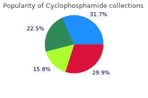
Beryllium (Be) is used in several forms: pure metal medicine 853 order 50 mg cyclophosphamide overnight delivery, beryllium oxide medicine hat college discount cyclophosphamide 50 mg on line, and as an alloy with copper treatment quad strain generic 50 mg cyclophosphamide overnight delivery, aluminum treatment erectile dysfunction discount 50 mg cyclophosphamide otc, or nickel medications in carry on luggage cyclophosphamide 50 mg without prescription. Beryllium oxide, beryllium metal and beryllium alloys are forms of Be present in the workplace with inhalation being the primary route of exposure. Therefore, a toxicological inhalation study using a murine model was performed in our laboratory in order to identify the toxic effects related to different particle sizes and chemical forms of Be. In this paper we describe the approach developed in order to reduce the potential for exposure, of technicians and students involved in the experiments, which includes specific attention to particle migration control through intensive housekeeping and systematic airborne and surface contamination. Considering the protection factor of 103 of a complete head cover used in this research, the average exposure level would be 0. Moreover, with the exception of one value, all average Be concentrations on surfaces were below the guideline level of 3 g/100 cm2 for Be contamination. Overall, employing a rigorous protective and preventive approach, will allow for future research to be conducted safely in this important field. Certain epidemiological data, but not all, suggest that soluble nickel exposure leads to increased respiratory cancer risk, but only with co-exposures to high levels of insoluble sulfidic or oxidic nickel. There is limited (although inconsistent) animal evidence that soluble nickel can act as a promoter, although it is unclear whether this mode of action is relevant to humans. Other in vitro studies have supported plausible non-genotoxic modes of action (cytotoxicity and induction of signal transduction pathways) for soluble nickel acting as a promoter. We considered whether the toxicological and mode-of-action data support the potential for soluble nickel to be either a complete carcinogen or a tumor promoter. We concluded that the weight of evidence does not clearly support a role for soluble nickel alone in carcinogenesis, and there is only limited evidence that it could act as a promoter. In the absence of specific toxicological data related to CdTe, regulatory agencies usually apply Cd criteria. However, it may be that CdThe has toxicological properties that are different from those of Cd, and it would therefore be useful to determine such data. Metals are known to influence oxidative stress resulting in patho-physiological conditions in aquatic organisms. Copper (Cu) and mercury (Hg) are the most diffused and hazardous organ specific environmental contaminants that exist in a wide variety of physical and chemical states, each endowed with unique characteristics of target organ specificity. Animals were collected from local fresh water ponds, acclimated to laboratory conditions for one week before the test animals were exposed to sublethal concentrations of Cu (0. The data indicate that Cu and Hg induced species-specific adaptive responses in these aquatic animals suggesting that the tissue antioxidants may further serve as surrogate markers of exposure to oxidant pollutants. Surveys show that the body burden of certain metals like cadmium or mercury is the consequence of active smoking habits. In a clinical trial 15 volunteers smoking about 15 cigarettes/day were included; this collective was randomly divided in two groups,blood and urine levels of cadmium,mercury and iron were measured under the influence of Cystus-Sud in a cross-over-design. Results: the enhanced levels of cadmium were reduced by 60% in blood and 45% in urine;with mercury the results were more indifferent between both groups. Our main finding was that Cystus-Sud significantly reduces the cadmium burden in smoking people,this confirms our pilot study in 8 volunteers in 1999. It has been known for decades that inhalation of sulfidic nickel ore refinery dust containing a mixture of soluble and insoluble nickel compounds can increase the risk of lung and nasal cancers in humans. What is less clear, however, is which specific chemical forms of nickel are associated with these cancers. To address the inconsistencies among studies of soluble nickel, we conducted a weight-of-evidence analysis of the relevant epidemiological, toxicological, and car- V. An extensive part of the Mexican territory has an inorganic arsenic (iAs) rich soil substrata, in addition anthropogenic activities such as mining and smelting process contribute to increase iAs levels in groundwater and soils leading to significant exposure to this metalloid. In Mexico, iAs exposure is mainly through drinking water coming from contaminated sources and affecting to 2 millions people. To achieve this objective, we conducted an exhaustive literature mining focused in geological data and its relevance to human health. Therefore biomarkers associated with health effects by chronic exposure to iAs have been actively investigated. In summary, due to the abundant presence of iAs in soil and water in Mexico, and its reported effects on human health, it is necessary to carry out systematic evaluations of the drinking water sources to identify all the high iAs areas and to develop surveillance program to detect early human effects. Increasing the dose above the current doses might show more significant biological changes in the lungs. In this study, we compared the toxicity and tissue distribution of the various types of As after repeated administration orally into the animal. The dosage of repeated administration orally into the monkey was determined based on the previous single administration study. Animals were administered with chemical in each group for 4 weeks except the sodium arsenite high-dosed group. We terminated the administration of As on day 6th, because observed died or abnormal positioned animals in the high dose sodium arsenite group. No significant change was not observed in the low-dose sodium arsenite and in the low- or high-dose arsenocholine administered animals during experimental period. Epidemiology studies have been unable to correlate chronic adverse lung effects associated with exposure to specific welding fumes generated from different processes. Generated fume was collected in the breathing zone of the animals, and particle size, morphology, and composition were determined. Bronchoalveolar lavage was done on days 1, 4, 8, 11, 22, and 43 after the last exposure to assess lung injury/inflammation and to recover lung phagocytes. Extremely high levels of alpha radioactivity in numerous domestic wells used for drinking water in the Fallon area were also found which could not be accounted for by naturally occurring uranium activity in the area and were subsequently determined to come from polonium-210 contamination. This study was designed to determine if consumption of drinking water from one or more sources high in one or more of these chemical from Churchill county Nevada induces oxidative stress in mice. Several sources of water samples were selected to provide representative ranges of naturally occurring levels of arsenic, tungsten and polonium-210. An in-depth chemical analysis of these water samples was conducted to confirm the concentration and diversity of the contaminants in these tests. Mice provided water high in tungsten but lower levels of polonium 210 and arsenic showed some increases in oxidative stress but not as significant as those high in all three. High tungsten levels are a common feature in the water samples which induce oxidative stress in this mouse model. Humans in Taiwan, Chile and Argentina exposed to inorganic arsenic in their drinking water have demonstrated elevated risks of lung cancer deaths. Arsenite-treated mice drank less water at all doses (significantly lower at 85, 10 and 1 ppm (66, 49 and 42 % respectively). Mouse lungs were analyzed for inorganic arsenic, monomethylated and dimethylated arsenicals by hydride generation atomic absorption spectroscopy. At 85 ppm, the total mean lung arsenical levels increased 14-fold and 131fold when compared to either the lowest non-control dose (0. Thimerosal (sodium ethylmercurithiosalicylate) is a potent antimicrobial agent extensively used as a preservative in pharmaceutical preparations such as immunogenic, ophthalmic and nasal products. The mechanism of the antimicrobial action of this organomercury compound has been ascribed to its strong affinity for sulfhydryl groups in cysteine residues of key microbial enzymes. In one set of experiments, the antifungal activity of these two compounds was tested using strains of both S. However, since thimerosal is cytotoxic at concentrations 1 M and an inhibitor of Pma1p at concentrations 10 M, its antifungal activity may not be solely dependent on its propensity to inhibit proton pumping function. Earlier publications found that a cytosolic pool of "unassembled" myosin light chain is released quickly from injured tissue while ischemia-induced acidosis and proteolytic degradation are possible mechanisms for subsequent dissociation of additional Myl3 from the myofibril enabling persistent release from injured myocytes. These data indicate that Myl3 is a sensitive and predictive biomarker of cardiac and skeletal muscle injury in veterinary species and adds value to a myopathy biomarker panel by extending the diagnostic window. The heavy metals in the human body cause damages to the health and the oligoelements are indispensable in small amounts for the life. Non invasive biomarkers are needed for monitoring and for that appropriate reference values are needed to be established. The study has been made in the municipality of Elche, province of Alicante Spain using children local population in the range of 3 to 12 years old that may considered a standard Spanish population from urban, rural, industrial areas. Values of reference for the heavy metal concentrations of the potentially toxic elements were established as the value in g of the element by gram of dry hair smaller than the 75 percentiles. For the oligoelements the reference value were established as the values between the percentiles 25 and 75. Myosin light chain-1 (Myl3) has been used as a biomarker of cardiac injury in humans to evaluate the severity and prognosis of patients with acute myocardial infarction and congestive heart failure. Current biomarkers of muscle injury in veterinary species appear quickly in blood and correlate well with injury. Myl3 has the added advantage of a wide diagnostic time window, remaining elevated in the blood for hours or days longer than many other markers. Myl3 gene expression and protein concentration are most abundant in heart and type-1 skeletal muscle. The integration of genomics data with traditional toxicology data remains a work in progress despite the positive strides taken by several members of industry. The end result is a robust tool with the potential to decrease the time and effort spent qualifying biomarkers and thus accelerating investigative compounds through the early phases of development. Further examples of data analysis for novel biomarkers of kidney, liver, and vascular injury will be discussed. The assays were validated to clinical laboratory standards that include the standard parameters of lowest detectable dose, normal range, dynamic range, imprecision, spiked recovery, linearity, cross reactivity and matrix interference. Results: - Assay development and validation efforts starting with multiple antibody reagents and standard calibrators lead to a robust panel of assays with the appropriate sensitivity and assay range. Multiplex assay performance was assessed with human urine samples from normal individuals and patients treated with the chemotherapeutics fluorouracil, cisplatin, gemcitabine, or the bisphosphonate, zoledronic acid. Histopathology confirmed that acute necrosis of proximal tubules was present in the high-dose group treated for 5 days. Multifocal accumulations of lymphocytes in the renal cortex associated with tubular basophilia were identified mainly in the mid- and high-dose groups treated for 14 days. Testicular toxicity is one of the target organ toxicities that are observed during preclinical development. Published literature discusses some biomarkers to monitor testicular changes following drug administration. This study focused on evaluating urinary creatine and gene expression changes as potential biomarkers of testicular toxicity. Serum Inhibin B levels will also be measured in this study to evaluate changes relative to the other markers. The ability to detect biologically relevant levels of toxicants and immune responses to toxicants presents a significant challenge. By coupling fluorescent emission from antibody-conjugated fluorophores to surface plasmons generated at the biosensor chip surface, fluorescent detection can be increased by 14-fold. This greatly expands the range of concentrations that can be detected for many analytes including toxicants, cytokines, enzymes, and other biomarkers. By utilizing a microarray format on a gold biosensor chip, we can simultaneously quantify hundreds to thousands of biomarkers reaching femtomolar sensitivity to generate unique signatures associated with disease states and toxicant exposure. Detected biomarkers can also include transcription factors, measures of enzyme specific activity, and other small molecules whose presence or change in concentration might be indicative of a disease state. The ability to define complex biological signatures may lead to earlier diagnosis and more effective treatments of disease and toxicant exposure. In conclusion, this novel and cost-effective blood preparation technique yields accurate results, requires less input than the traditional method, and can also be used in preclinical toxicology and pharmacology studies to identify novel biomarkers. The kidney is one of the main targets of drug-induced toxicity, but early detection of renal damage is often difficult. As part of the InnoMed PredTox project, a collaborative effort by 15 pharmaceutical companies, 2 small- and mid-sized enterprises and 3 universities aimed at assessing the value of combining omics technologies with conventional toxicology methods for improved safety assessment, we evaluated the performance of a panel of novel kidney biomarkers in preclinical toxicity studies. Rats (Han Wistar) were treated with either the reference nephrotoxin gentamicin or one of several proprietary compounds that were dropped from drug development in part due to renal toxicity. Changes in gene/protein expression generally correlated well with renal histopathological alterations and were frequently detected at earlier timepoints or at lower doses than traditional clinical parameters. Overexpression of Kim-1 was often one of the earliest responses and might be seen as the most sensitive tissue marker of kidney injury. Urinary Kim-1 and clusterin reflected changes in gene/protein expression and histopathological alterations in the target organ, confirming clusterin and Kim-1 as early and sensitive, non-invasive marker of renal injury. In contrast, urinary lipocalin-2 was found to be less sensitive and not specific for kidney toxicity. Chronic low-level lead toxicity takes a silent toll on those it effects, driving cognitive and behavioral alterations that are not apparent for years after exposure. Here genomic and proteomic approaches are used in parallel to identify gene expression and functional protein interaction responses to low-level lead intoxication. The result is a set of candidate molecular biomarkers of chronic low-level lead intoxication. Initial proteomic screens utilized a modified two-hybrid proteomic system to identify a number of potential in vivo protein-protein interactions disrupted by the neurotoxin lead (Pb2+). In the second phase of this project the proteomic set of molecular markers was compared with microarray data, global gene expression patterns observed in offspring of time-pregnant Long Evans rats subject to chronic low-level lead exposure vs. Both genomic and proteomic methods described are subject to artifacts, but when the data from each method is overlaid, elements present in both data sets identify a subset of molecular biomarkers. To date we know of no reliable biomarkers for low-level lead toxicity in man, nor is there a satisfying model for how chronic low-level lead toxicity effects the neuronal systems of mammals. This work provides a glimpse at these mechanisms, identifies a candidate protein network of biomarkers for chronic low-level lead exposure, and describes a novel platform for de novo biomarker discovery for poorly characterized toxicant/organism systems. Blood is an easily accessible tissue that can be used to identify biomarkers for a wide range of tissue injuries using transcriptomic profiling.
50 mg cyclophosphamide for sale. #TomorrowsDiscoveries: Detecting HIV prior to symptoms—Dr. Gabe Kelen.
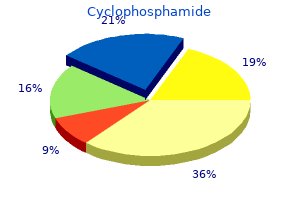
References
- Norrsell H, Eliasson T, Augustinsson LE, Mannheimer C: [Spinal cord stimulation in angina pectorisowhat is the current situation?] Lakartidningen 1999;96:1430-1432, 1435-1437.
- Dietrich CF, Sahai AV, D'Onofrio M, et al. Differential diagnosis of small solid pancreatic lesions. Gastrointest Endosc. 2016;84(6):933-940.
- Burke NJ, Joseph G, Pasick RJ, Barker JC. Theorizing social context: rethinking behavioral theory. Health Educ Behav 2009; 36(5 Suppl):55S-70S. 8.
- Whooley MA, Avins AL, Miranda J, et al: Case-finding instruments for depression. Two questions are as good as many, J Gen Intern Med 12(7):439n445, 1997.
- Taylor LM, Park TC, Edwards JM, et al: Acute disruption of polytetrafluoroethylene grafts adjacent to axillary anastomoses: a complication of axillofemoral grafting, J Vasc Surg 20:520, 1994.
- Moravan M, Segal BM. Treatment of CNS sarcoidosis with infliximab and mycophenolate mofetil. Neurology. 2009;72(4): 337-340.
- Nonaka I, Sunohara N, Satoyoshi E, Terasawa K, Yonemoto K. Autosomal recessive distal muscular dystrophy: A comparative study with distal myopathy with rimmed vacuole formation. Ann Neurol 1985;17(1):51-59.
- Swartz WM, Banis JC, Newton ED, et al. The osteocutaneous scapular flap for mandibular and maxillary reconstruction. Plast Reconstr Surg 1986;77:530.