- info@careermakers.edu.np
- +977 1 4423870
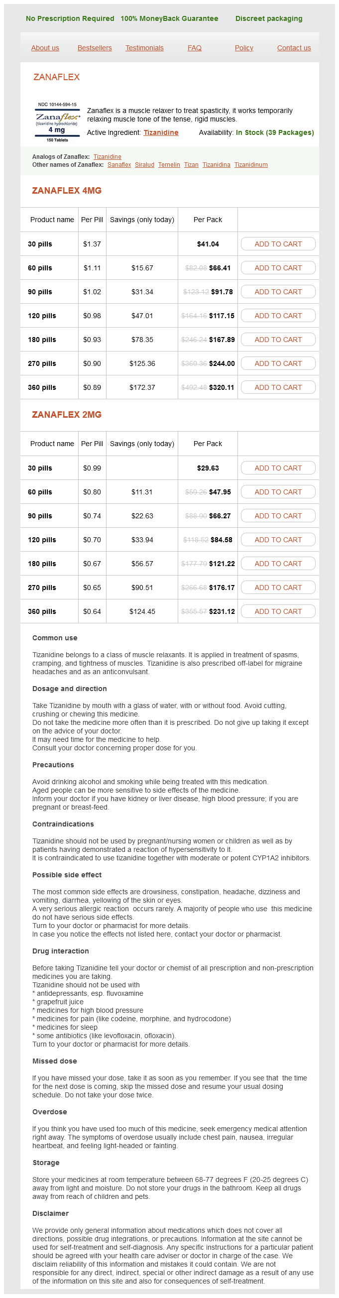
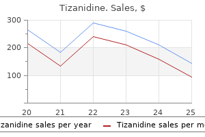
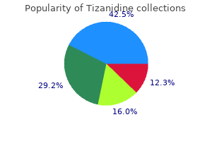
Tizanidine
Brent Grady, RN, CCRN, CEN
- Flight Nurse
- Loyola Hospital
- Maywood, IL
This autoimmune response Constridive Pericarditis Constrictive pericarditis is a disease that is being encountered with increasing frequency muscle relaxant tramadol buy tizanidine 4 mg without prescription. The most frequent inciting factor is postoperative bleeding associated with cardiac surgery spasms pelvic area purchase tizanidine 4 mg on line, especially coronary revascularization procedures spasms left upper abdomen 4 mg tizanidine. Frontal (left) and lateral posterior evagination (right) radiographs show left retrocardiac density (arrow) on the frontal view and large (arrowheads) of left ventricular contour on the lateral view muscle relaxant sciatica order tizanidine 2 mg without a prescription. Recognition of pericardial calcification supports or may initially suggest the diagnosis of constrictive pericarditis muscle relaxant 4211 order tizanidine 2 mg with amex. Cardiomegaly is usually indicative of acute infarction in a patient with history of previous infarctions. Signs of complication of acute myocardial infarction Intractable pulmonary edema may occur with papillary muscle rupture (mitral regurgitation) or ventricular septal rupture (left to right shunt). Abnormal cardiac contour may be a sign of true (bulge of the anterolateral or apical regions; see. A signpost pointing to the aortic valve is present in this disease, consisting of enlargement of the ascending aorta, aortic knob, and usually the descending thoracic aorta. As opposed to aortic stenosis, the enlargement of the thoracic aorta involves the aortic knob as well as the ascending aorta. Uremic pericardial disease may also eventuate in constrictive pericarditis, but usually this disease produces an effusive/constrictive type of pericardial disease. In Third World countries, tuberculosis continues to be a major cause of constrictive pericarditis. The plain radiograph is frequently but not always abnormal in patients with hemody namically significant constrictive pericarditis. Because of pericardial constriction there is restriction to left atrial emptying during diastole, with a subsequent rise in left atrial and pulmonary venous pressures. The rise in pulmonary venous pressure is reflected on the Consequently, "big heart" heart disease with the aortic signpost is indicative of aortic regurgitation. Since this is a volume overload lesion, the extent of the increase in volume of the heart is related to the severity and the duration of aortic regurgitation. For most of the course of aortic regurgitation, the pulmonary vascularity is normal. Frontal (left) and lateral (right) radiographs show grade I pulmonary ventricular hypertension and flattened right cardiac contour (arrows), which are char acteristics of constrictive pericarditis. Lateral view demonstrates calcification (arrowhead) in the posterior interventricular groove. The calcification involves the atrioventricular (right) radiographs (arrows) and demonstrate the interven (arrowheads) grooves. The giant left atrium can be associated with either mitral stenosis or regurgitation but is more frequently caused by the latter. The right border of the left atrium may even extend beyond 30-9 to 30-11, 30-18, and 30-19) and sometimes signs of right-sided chamber enlargement 30-10 and 30-11). In the presence of isolated mitral regurgitation the ascending aorta is relatively small. Consequently, recognition of prominence of the ascending aorta in a patient with isolated mitral valve disease raises the prospect of associated aortic valve disease. The left atrial appendage is generally enlarged in patients who have a rheumatic etiology of the mitral regurgitation. On the other hand, the left atrial appendage is frequently not enlarged in patients who otherwise have left atrial enlargement of nonrheumatic etiology. Frontal radiograph shows marked cardiomegaly with displacement of the ventricu lar contour laterally and caudally, indicating left ventricu lar enlargement. Concavity Pulmonary venous hypertension Normal heart size or mild cardiomegaly Left atrial enlargement may be discernible. Calcification of the cardiac margin, especially the atrioventricular and interventricular grooves. The plain radiograph may be useful in assessing the severity of mitral regurgitation. Because this is a volume overload lesion, the overall heart size may be a reasonable indicator of the severity of regurgitation. Likewise, the overall heart size may be of some prognostic use in patients undergoing mitral valve replacement. In general, patients with lesser degrees of cardiomegaly demonstrate a greater 5-year survival rate after replacement of the mitral valve (Table 30-14). The features of this lesion are diminished pulmonary vascularity, marked car diomegaly, and right atrial and right ventricular enlarge ment. The severe enlargement of the right-sided chamber produced the "wall-to-wall heart. Signs of right atrial enlargement are frequently dubious and not sharply discriminated from normal. Indeed, there must be substantial right atrial enlargement before it is possible to recognize its occurrence. In general, the best sign of right atrial enlargement is elongation of the right atrial border. The radiographic signs of tricuspid regurgitation are normal or perhaps reduced prominence of the pulmonary vascularity, cardiomegaly, right atrial enlargement, and occasionally signs of superior vena caval and especially inferior vena caval enlargement. Cardiomegaly, with the signpost of right atrial enlargement, would indicate the likely diagnosis of tricuspid regurgitation. The cardiac contour in patients with tricuspid regurgitation may be similar to that of congestive cardiomyopathy and pericardia! The most extreme cardiomegaly is seen with severe tricuspid regurgitation of long duration; it can cause the "wall-to-wall" heart. Congestive Cardiomyopathy the radiographic appearance in congestive cardiomyopathy is relatively nonspecific. Characteristically, the cardiomegaly exists without the presence of signposts to the aortic, mitral, or tricuspid valve. Consequently, substantial cardiomegaly ("big heart" heart disease), without radiographic signposts, should raise the diagnostic consideration of congestive cardiomyopathy. At the current time, the most frequent cause of congestive or dilated cardiomyopathy is ischemic heart disease. However, from a strict classification point of view, ischemic heart disease should not be considered as part of the group of congestive cardiomyopathies. Frontal radiograph shows biventricular enlargement and mild pulmonary ventricular hypertension (grade I). Enlarge ment of the left ventricle is indicated by a vector of ven tricular enlargement directed laterally and caudally on the frontal view. Right ventricle enlargement is indicated by the prominent convexity of the upper left cardiac border on the frontal view. Myocardial Disease as a dilated cardiomyopathy without known etiologic identification (Table 30-16). A specific appearance providing a diagnosis of pericardial effusion is relatively infrequent in this entity. The so-called water-bottle appearance of the heart is nonspecific and difficult to recognize. The "fat pad" sign seen on the lateral radiograph does permit identification but occurs in only a few patients. This consists of a lesser density at the periphery of the cardiac contour compared to the central portion of the cardiac contour. The cause of this varying density is that the x-ray beam encounters only fluid toward the periphery of the pericardial effusion, while in the center of the pericardial effusion the radiographic beam must pass through both water anteriorly and the cardiac substance more centrally. With the frequent use of echocardiography, large be pericardial effusions are being encountered less frequently. The presence of any degree of pericardial effusion easily recognized by echocardiography (Table 30-17). It has been assumed that the presence Paracardiac Masses Enlargement of the cardiac contour may not always be indicative of cardiac enlargement itself or pericardial effusion. One must also consider the infrequent possibility that the enlargement represents a cardiac or paracardiac mass. Such consideration should be prompted by recognition of an unusual cardiac contour. Frontal radiograph shows moderate cardiomegaly with biven tricular but no discernible left atrial enlargement. A stripe of water density (arrow) separates two fat layers on the outer surface of the parietal pericardium and beneath the visceral pericardium. The differential diagnosis of pulmonary hypertension should lead to a systematic organization of the diagnostic possibilities. There are a number of causes of enlarge ment of the main pulmonary artery (Table 30-18). Enlargement of the main pulmonary artery segment is the main indicator of pulmonary arterial hypertension (1) pulmonary arterial hypertension resulting (2) pulmonary arterial hypertension resulting from left-to-right shunts resulting in pulmonary arteriolar disease (arteriolopathy); (3) pulmonary arterial hypertension resulting from obliteration of the pulmonary vascular bed from chronic lung disease; (4) pulmonary arterial hypertension resulting from obliteration of the pulmonary vascular bed as a consequence of pulmonary embolic disease or schistosomiasis; and (5) primary pulmonary hypertension. Radiographic signs that permit the differential diagnosis of the various causes include recognition of the following: 1. Signs of chronic lung disease such as chronic obstructive pulmonary disease or interstitial lung disease would indicate this as the etiology. Varying cardiac density Moderate to severe enlargement of cardiac silhouette Associated pleural effusion is not uncommon Specific features, such as "fat pad" and/or "variable density' signs, are infrequently evident. Asymmetric pulmonary vascularity or signs of pulmonary scarring might indicate the presence of chronic thromboembolic disease. The left atrial appendage region is considered to be the region immediately adjacent to and below the left bronchus. This is in contradistinction to the region of the main pulmonary artery segment, which is above the left bronchus. The two normal structures that reside within this area are the left atrial appendage and the right ventricular outflow tract; the left atrium is situated posterior to the right ventricular outflow region. The pericardium covers the left atrial appendage and the right ventricular outflow tract in this region as in other parts of the heart. Frontal view shows the markedly enlarged main (arrow) and right pulmonary arterial segments. There is calcification (arrow) in the pulmonary arteries consistent with a systemic arterial pressure level in the pulmonary circulation. The abnormally positioned structures that can lie within this site are a juxtaposed right atrial appendage and a transposed ascending aorta with the associated inverted right ventricular outflow region. Enlargement of the Left Lower Cardiac Border-Ventricular Region Enlargement along the lower left cardiac border in the region of the ventricles is most frequently caused by enlargement of either the right or the left ventricle. An abnormal convexity evagination within this region has a Enlargement of the Right Heart Border Enlargement of the right heart contour in the frontal view is generally ascribed to right atrial enlargement. Complete absence causes a shift of the heart to the left without a shift of the mediastinum (note the central position of the trachea) and a prominent convexity of the upper left cardiac margin. Lung (arrow) is insinu ated between the aorta and pulmonary artery, indicating an absence of the pericardium at this site. Partial absence causes a prominent convexity (arrowhead) of the mid-left cardiac margin, especially in the region of the left atrial appendage segment. Aortic annular calcification, a circular calcification in the region of the aortic valve. Extension of this calcification into the region of the conducting system can produce complete heart failure. Calcification Cardiac Calcification Calcification in the central cardiovascular structures is frequent and is an important diagnostic sign. In a few instances, calcification of a specific shape and location is pathognomonic for a disease. The various cardiovascular calcifications include the following: of the aortic valve of sufficient density and extent to be visualized on the radiograph. This is most frequently observed on the right anterolateral margin of the ascending aorta in elderly individuals, especially in the presence of aortic valve disease. It is located usually in the interventricular or atrioventricular grooves of the heart. Unusual sites of calcification may represent intracardiac tumor (left atrial myxoma), pericardia! This is the most frequent site where coronary arterial calcification is observed on the frontal radiograph. Assess ment of the plain radiograph can usually provide only a notion of the generic type of congenital heart lesion rather than a dear indication of specific lesions. An approach that is cognizant of the realistic insights possible from the plain radiograph must be pursued. Such an approach should be based on the observations on the radiograph that can be made with some degree of certitude and in which there is minimal ambiguity. The current classification system depends on a few clini cal observations and a few findings on the radiograph that can be made with reasonable reliability. A fifth group consists of patients with primarily pulmonary venous congestion 31-2). Therefore, when interpreting the chest x-ray, the physician attempts to decide which class or category of congenital heart lesions exists. The decision on the specific lesion is usually based on the statistical frequency of a par ticular cardiac lesion within one or more groups. Based on the clinical and radiographic findings, there are five groups of congenital heart lesions.
Prominent upper left cardiac border on frontal view is caused by dilatation of the right ventricular outflow region muscle relaxant and tylenol 3 buy tizanidine with visa. Lateral view shows posterior displacement of the left bronchus (arrow) by the enlarged left atrium and obliteration of the retrocardiac space by right ventricular enlargement spasms perineum generic 2 mg tizanidine free shipping. On the posteroanterior view spasms in upper abdomen buy generic tizanidine 2 mg on line, leftward and downward bulging of the right posteroanterior radiograph muscle relaxant 750 order tizanidine cheap online. A rough rule is that a right atrial border exceeding 60% in length of the mediastinal cardiovascular shadow is a sign of substantial right atrial enlargement spasms synonyms buy 4 mg tizanidine with mastercard. Severe right atrial enlargement is evident by the elongation of the right atrial shadow. The length of the right atrial border exceeds 600/o of the height of the mediastinal cardiovascular structures. The ventricular contour is enlarged along a left inferolateral vector, causing the apex to droop over the left hemidiaphragm. Concavity along the upper left cardiac border indicates that the right ventricle is not enlarged. The vector of enlargement of the ventricle is directly lateral, indicating right ventricular enlargement. On the posteroanterior view, the left border of the heart monary arterial hypertension is indicated by pulmonary arterial enlargement. This schema obviously works well is enlarged directly laterally or laterally and slightly superiorly. In some instances, this causes the apex to be displaced superiorly ("upward tipped apex";. On the lateral view, the retrosternal space is encroached upon by the enlarged right ventricle. Right ventricular enlargement is inferred by contact of the right heart border over greater than one third of the sternal length. A prominent convexity to the anterior border rather than the usual straight surface is an early sign of right ventricular enlargement. Signposts for Cardiac Valvular Lesions There are three signposts on the thoracic radiograph that direct attention to a certain cardiac valve: 1. The vector of enlargement of the ventricle is leftward and cranial, causing uplifting of the apex in relation to the diaphragm. Frontal radiograph demonstrates heavy calcification (left) shows a nearly normal appearance except for enlargement of the ascending aorta. Of course, a specific cardiac lesion does not always cause typical features because of other associated abnormalities or because the lesion is very mild or has been present for insufficient time to alter the cardiac morphology to a degree discernible on the thoracic radiograph. The schema can be briefly described by considering the chest x-ray that shows a normal heart size or mild cardio megaly in a patient with significant heart disease. If the left atrial signpost is present, then attention is directed to the mitral valve. The schema for a patient with substantial cardiomegaly proceeds along the following path. The big heart suggests that there is either a volume overload lesion (valvular regur gitation) or myocardial failure or pericardia! High output states are certainly a volume overload and can cause substantial cardiomegaly, but sometimes they cause only mild cardiomegaly. If left atrial enlargement is noted, then the signpost points to mitral regurgitation. The diagnosis should be either mitral valvular stenosis or resistance to left atrial emptying. Ifthe ascending aorta is enlarged, then this signpost points to the aortic valve, indicating aortic stenosis. Systemic hypertension can produce a similar appearance, although it usually causes enlargement of the entire thoracic aorta rather than the ascending aorta alone. If no signposts are present, then the diagnosis is unlikely to be a valvular lesion. However, even these latter diseases sometimes induce left atrial enlargement, as stated above. Mitral regurgitation causing cardiomegaly and left ventricular and left atrial enlargement. The sole abnormality in this 40-year-old subject is enlarge ment of the ascending aorta (arrows). If the right atrium is enlarged, then this signpost points to tricuspid regurgitation. Acquired pulmonic regurgitation is rare, except as a consequence of operation for right ventricular outflow obstruction, and is not considered in this schema. If no signposts are present, then the favored diagnostic considerations are congestive (dilated) cardiomyopathy or pericardia! Consequently, aortic stenosis, for much of its natural history, is a disease that is clearly "small heart" heart disease. The pulmonary vascularity is also generally normal for much of the course of aortic stenosis. Mitral Stenosis the features of a thoracic and radiograph are frequently diagnostic for mitral stenosis. Frontal thoracic radiograph demonstrates left atrial and right ventricular enlargement. Left atrial enlargement is indicated by right retrocardiac double density (arrow) on the frontal view. There is pul monary arterial hypertension as shown by enlargement of the main and central pulmonary arteries. Right ventricular enlargement is indicated by lateral displacement of the ventricular mar gin (apex uplifted) on the frontal view. Likewise, the radiograph provides considerable insight into the severity of mitral stenosis. Hypertrophic Cardiomyopathy the chest radiograph is neither specific nor sensitive for the diagnosis of hypertrophic cardiomyopathy. More than 50% of patients with hypertrophic cardiomyopathy have a normal chest x-ray. In a few patients there is some abnormality of the chest x-ray, which is usually relatively vague and not particularly indicative of this disease. Approximately 30% of patients with symptomatic hypertrophic cardiomyopathy have associated mitral regurgitation. Because of the mitral regurgitation, there again is a proclivity to left atrial enlargement. Frontal view shows borderline cardiomegaly and prominence of the entire thoracic aorta. This focal enlargement is a consequence of extreme enlargement of the upper or outflow portion of the ventricular septum (Table 30-9). In the obstructive form (subaortic stenosis), ascending aortic enlargement is infrequent. During the early stage of restrictive cardiomyopathy the cardiac size is within normal limits. Restridive Cardiomyopathy Restrictive cardiomyopathy is a relatively rare disease that may occur in an idiopathic form or may be the form of cardiomyopathy that is a consequence of various infiltrative 30-26). Enlargement of the left atrial appendage is frequent and suggests a rheumatic etiology. Right ventricular enlargement indicates some degree of pulmonary arterial hypertension or associated tricuspid regurgitation. Right ventricular enlargement in the absence of prominence of the main pulmonary artery suggests associated tricuspid regurgitation. The ascending aorta and aortic arch are usually inconspicuous in isolated mitral stenosis. Even slight enlargement of the thoracic aorta raises the question of associated aortic valve disease. Frontal (left) and lateral (right) radiographs show inter stitial pulmonary edema and left atrial and right ventricular enlargement. However, in many patients there is evidence of an abnormal cardiac configuration, especially an abnormal evagination along the midportion of the left cardiac border or in the region of the cardiac apex. Note alveolar filling in perihilar regions and lower lobes with normal heart size. Consequently, an abnormal contour or a double density localized to these sites should raise the consideration of this diagnosis. Other plain radiographic signs of a false aneurysm are an aneurysm that is extremely large with a prominent projection off the posterior or diaphragmatic surface of the heart. The initial chest x-ray film in acute myocardial infarction: prediction of early and late mortality and survival. Partial rupture of the papillary muscle, resulting in less severe mitral regurgitation, may produce a moderate degree of mitral regurgitation and less severe or even no evidence of pulmonary edema. The dramatic radiographic findings in acute papillary muscle rupture are pulmonary edema with little increase in left atrial size or cardiomegaly. Postinfarction rupture of the ventricular septum may produce a radiographic appearance very similar to that of acute mitral regurgitation. The radiographic signs of acute ventricular septal defect include an increase in the prominence of the pulmonary arteries. Abnormal evagination of left cardiac border (arrowheads) is typical for an aneurysm involving the anterolateral and/or apical segment of the left ventricle. Lateral view dem onstrates an anterior double density (arrows), characteristic of an anterolateral aneurysm. Lat eral thoracic radiograph shows calcification the anteroapical region of the left ventricle. Chest radiographs in this syndrome demonstrate an increase in cardiac size as a consequence of the pericardia! Group I: left-to-right shunts Noncyanotic: Sometimes symptoms of pulmonary con gestion or congestive heart failure. Hilar vessels are small and segmental pulmonary arteries are hardly visible, espe cially in the upper lobes. Vector of enlargement of the apex of the heart is directly lateral, indicating right ven tricular enlargement. Pulmonary arterial overcirculation is shown by large hilar and segmental pulmonary arteries. Note pulmonary arte rial overcirculation in the presence of cyanosis and cardio megaly. It is frequently difficult to distinguish between normal and diminished pulmonary vascularity. The criteria that place a patient within this category are for the most part dependent on the clinical recognition of the absence of cyanosis with the sub sequent demonstration on the chest radiograph of increased pulmonary arterial vascularity. Consequently, the major observation on the radiograph in terms of pulmonary vas cularity in the cyanotic patient is to determine whether the pulmonary vascularity is increased. Normal or diminished pulmonary vascularity in a patient with cyanosis indicates that the lesion produces a right-to-left shunt. Increased pul monary vascularity in a cyanotic patient indicates that there is an admixture lesion; the cyanosis is indicative of right-to left shunting, and increased pulmonary vascularity is a sign of left-to-right shunting. The degree of cardiomegaly is usually in proportion to the increase in pulmonary vascularity. Consequently, there is frequently cardiomegaly, and this cardiomegaly should in general be in proportion to the prominence of the pulmonary vascularity. Note pulmonary arterial overcir evidenced by shunt vessels and prominent hilar vessels. Left atrial enlargement produces impression on and dis placement of the barium-filled esophagus, as shown on the lateral view. The prominent aortic arch (arrow) and descending aorta are diagnostic signs of pat ent ductus arteriosus. On the lateral view, the enlarged left atrium causes posterior displacement of the left bron chus (arrowhead). In infants, prominence of the aortic arch may be difficult to recognize, so this signpost may not always be available. The severe volume overload with large left-to-right shunts causes pul monary venous congestion or pulmonary edema in addition to pulmonary arterial overcirculation. In indi viduals with large left-to-right shunts, there should also be substantial cardiomegaly. When cardiomegaly exists out of proportion to the pulmo nary arterial vascularity, then one must consider a number of possibilities. Another consideration is the coexistence of additional cardiac lesions, such as pri mary myocardial disease or coarctation of the aorta. Two signposts can be used to help distinguish among the various types of left-to-right shunts. Large-volume left to-right shunt causing pulmonary edema, severe pulmo nary arterial overcirculation, and cardiomegaly. Indistinct hilar and segmental arteries on the right side are caused by interstitial edema. Note pulmonary oligemia with more diminished vascularity on the left, especially the left upper lobe. Normal heart size and concave pulmonary artery segment are characteristic features in the infant. The patient with tricuspid atresia with a large atrial septal defect demonstrates little or no cardio megaly. On the other hand, the patient with tricuspid atresia with a restrictive atrial septal defect expe riences substantial right atrial enlargement, which results in cardiomegaly. The remaining diagnostic considerations are, for the most part, variants of tetralogy of Fallot.
Magnetic resonance-defined periportal steatosis following intraportal islet transplantation: a functional footprint of islet graft survival Percutaneous transhepatic pancreatic islet cell transplantation in type 1 diabetes mellitus: radiologic aspects muscle relaxant alcoholism cheap tizanidine master card. Long-term survival following simultaneous kidney-pancreas transplantation versus kidney transplantation alone in patients with type 1 diabetes mellitus and renal failure muscle relaxant with painkiller buy generic tizanidine 2 mg online. Islet transplantation using donors after cardiac death: report of the Japan Islet Transplantation Registry muscle relaxant carisoprodol order tizanidine discount. Influence of Pancreas Preservation on Human Islet Isolation Outcomes: Impact of the Two-Layer Method muscle relaxant vitamins discount tizanidine 4 mg buy online. Pancreatic islet transplantation after upper abdominal exenteration and liver replacement spasms rib cage area discount tizanidine 2 mg amex. Introduction During the past decade, simultaneous pancreas kidney transplantation has been widely accepted as the most effective way to achieve normoglycemia in patients with type 1 diabetes and end-stage renal disease. Unfortunately, pancreas transplantation has been associated with the highest surgical complication rate of all the routinely performed organ transplant procedures (except for small intestinal or multivisceral transplantation) and the risk of pancreas graft loss from surgical complications (technical failures) is higher than from immunological reasons. Compared to patients with diabetes who receive a kidney alone, the addition of a pancreas improves long-term patient and kidney graft survival. Recipients of a pancreas-after-kidney transplant or a pancreas transplant alone have an average 1-year pancreas graft survival rate of 78-83%. In this chapter we will thoroughly describe many aspects of this complex transplantation procedure including: the history of pancreas transplantation Indications for pancreas transplantation Surgical aspects of pancreas graft procurement from the deceased donor Current surgical techniques for pancreas transplantation Immunosuppressive regimens 388 - Understanding the Complexities of Kidney Transplantation Postoperative care of pancreas transplant recipients Complications of pancreas transplantation Long term results of pancreas transplantation 2. Only 12 cases were done between 1967 till 1973 at the same center but almost all of them rejected before the first year after the operation. Besides the rejection and vascular problems, dealing with pancreas exocrine secretion was complicated this type of procedure from the early days. By the mid-70s three different techniques were in use: enteric drainage, urinary drainage (into the ureter or directly into the bladder and duct injection. In 1984, Starzl et al (Starzl et al,1984) reintroduced the technique of enteric drainage as originally described by Lillehei which is now is the routine procedure in most pancreas transplant centers. Indications for pancreas transplantation Patients with type 1 or insulin dependent type 2 diabetes mellitus are eligible for pancreas transplantation when they have any major complications of their disease, but because of complications of this type of surgery and need for lifetime immunosuppression therapy and frequent interventional surveillance (such as protocol biopsies), both the clinicians and the Kidney-Pancreas Transplantation 389 patients should be aware of those conditions that really might benefit from pancreas transplantation. Most common indications are as follows: diabetic nephropathy, retinopathy, neuropathy, disabling or life threatening hypoglycemic unawareness or incapacitating emotional or clinical problems associated with insulin therapy (White et al, 2009). Some of the rare indications (which are not accepted by all transplant surgeons) are diabetic complications after total pancreatectomy, presence of other autoimmune diseases, insulin allergy or resistance to subcutaneous insulin. Unfortunately, most patients with chronic diabetes who develop these complications have major comorbidities such as obesity, cardiovascular, cerebrovascular or peripheral vascular diseases, diabetic gastropathy, and vascular or neuropathic diabetic foot. These patients are most suitable for pancreatic islet cell transplantation which is very simpler and has fewer and more minor complications that the relatively complex surgical procedure of whole pancreas transplantation. When coordinate logistics available, these operation using kidney graft from the living donor and pancreas graft from a deceased donor may be performed at the same time and at the same center. Surgical aspects of pancreas graft procurement from the deceased donor Not all deceased donors are suitable for pancreas graft procurement. Donor hyperglycemia is common because of stress condition and use of high dose corticosteroids and not a contraindication for use of the pancreas, although it may contribute as a minor risk factor for long-term graft loss (Gores, et al, 1992). Most surgeons only select younger non-obese donors (age 10-50 years, weight 30-100 kg) who are hemodynamically stable without need to high doses of vasopressors. Also a cardiocerebrovascular cause of brain death and massive volume resuscitation are other risk factors for postoperative complications in the recipients (Troppmann C, 2004). Because pancreas transplantation is not life saving, procurement of other donor organs are more important. If the surgeon considers that the pancreas is not suitable for transplantation it may be used for research or pancreas islet cell transplantation (Shapiro, et al, 2000). The procedure starts with a long midline incision from suprasternal notch to symphysis pubis area usually by the liver team. All intrathoracic and abdominal organs are evaluated systematically to rule out any suspicious lesion. First, all usual dissections for controlling supraceliac and infrarenal aorta, arc of thoracic aorta and superior or inferior mesenteric vein should be performed, so in case that patient becomes unstable the surgeon can rapidly proceed with cold perfusion of the organs for their safe retrieval. The right colon is completely mobilized from retroperitoneum and then an extended Kocher maneuver is done. All ligaments of the liver are transected and then arterial anatomy of the liver and pancreas is evaluated by palpating the hepatic artery pulsation in the hepatodudenal portion of lesser omentum. The surgeon should have complete knowledge of hepatic artery abnormalities and possibility of existence of a right accessory or right replaced hepatic artery that originate from superior mesenteric artery. In rare cases the entire hepatic artery are originated from superior mesenteric artery. With novel microsurgical techniques none of these anomalies is considered as a contraindication for concomitant liver, pancreas or small intestinal harvesting from a deceased donor. The common bile duct is divided and infrarenal aorta and superior or inferior mesenteric vein are cannulated at the next stage. Supraceliac aorta is clamped 3-5 minutes after systemic heparinization and the heart team also clamps the aortic arc and cold perfusion is started. After removing the heart and lungs, liver, pancreas and small intestine usually are procured en bloc and the remaining dissection may be performed in the bench procedure. The gastroduodenal artery is divided and suture ligated and the splenic artery is divided close to its origin and marked by a nonabsorbable 6-0 suture for future identification. In back table or bench procedure, all excessive fat tissue and spleen should be removed and the origin of mesentery and all small arterial and venous branches in the inferior border of pancreas is reinforced again for future hemostasis. Duodenum is shortened again and reinforced in both distal and proximal side by non-absorbable sutures. Arterial reconstruction is performed by anastomosis of the donor iliac Y-graft, external and internal iliac branches to the graft superior mesenteric and splenic artery, respectively. We also recommend using a small segment of donor left gastric or inferior mesenteric artery for reperfusion of gastroduodenal artery for better circulation of duodenum and head of pancreas to prevent future frequent duodenal ulcers in the graft. During kidney transplant procedure, the pancreas team prepares the pancreas graft for transplantation. Usually an intraperitoneal approach is used by a long midline incision and the kidney graft is transplanted by standard technique to left iliac fossa (renal artery to internal or external iliac artery and then renal vein to external iliac vein and at last ureter to the bladder or native ureter as described in other chapters of this book). Use of right side for pancreas transplantation is recommended due to more superficial iliac artery position in this side, which makes arterial anastomosis easier. Except for a few minor changes in arterial reconstruction technique (such as reperfusion of gastroduodenal artery or changing the site of arterial inflow), there is no significant change in the arterial reconstruction technique during these era. In our center we use the recipient superior mesenteric vein at the base of mesentery below the transverse mesocolon for venous outflow(portal drainage) and right common iliac artery for arterial inflow to the donor iliac Y-graft. Those surgeons that prefer to use systemic venous drainage use the right external or common iliac vein as the venous outflow, perfectly as the same manner that they used 392 Understanding the Complexities of Kidney Transplantation external iliac artery and vein for kidney transplantation. After completing the arterial and venous anastomoses, the graft is reperfused and complete hemostasis is done. Usually despite every effort for complete hemostasis during back table preparing of the pancreas graft, there is brisk bleeding around the borders of the graft at the time of reperfusion and crystalloid and blood product replacement should be used to prevent hypotension and subsequent damage to the kidney and pancreas grafts. The next step is anastomosis of the graft duodenum to the recipient jejunum (enteric drainage) or bladder (bladder drainage). When portal drainage is chosen, the pancreas head should be directed cephalad and use of bladder drainage is almost impossible and vice versa. Also if portal drainage is used we need a rather longer donor common iliac artery or even an extension graft for arterial reconstruction of the graft. Drains are inserted at the end of operation around the kidney and pancreas graft separately to monitor for postoperative bleeding and leakages. We prefer to perform the kidney transplant operation retroperitoneally in the left iliac fossa and at the end use the peritoneum to hide the kidney to be able to monitor the kidney graft complications such a urine leakage and lymphocele s1eparately from the pancreas graft. Enteric drainage also has many complications (at least risk of enteric contamination) that may be devastating in case of duodenal necrosis and gastrointestinal leakage. Several options are available for enteric drainage: side-to-side duodenojejunostomy, or duodenojejunostomy with a Roux-en-Y limb and duodenoduodenostomy (Hummel et al, 2008). The site of duodenojejunostomy (distance from the Treitz ligament) and its length are different between authors. Although duodenoduodenostomy complications are more Kidney-Pancreas Transplantation 393 dangerous but endoscopic biopsy and hemostasis of duodenal ulcers of the transplanted pancreas-duodenum complex will be made feasible by this technique. Systemic venous drainage is technically less demanding and used with less difficulty and more frequently by those surgeons who are familiar with kidney transplantation technique. In the early days of pancreas transplantation in the University of Minnesota, portal venous drainage was used for the pancreas graft venous effluent only in seven cases. Systemic hyperinsulinemia caused by systemic drainage first was showed by Diem et al (Diem et al, 1990). Some centers now use extra- or retroperitoneal approaches for better accessibility of the pancreas graft for postoperative routine percutaneous biopsies and easier arterial anastomosis and some of them suggest using an en bloc kidney-pancreas transplantation from the same donor. The technique of retroperitoneal pancreas transplantation with portalenteric drainage was first described by Boggi et al in 2005 (Boggi et al, 2005). This method may be used in patient with severe intraperitoneal adhesions due to multiple previous abdominal surgeries and also for pancreas retransplant. Kahn et al described the same technique by systemic venous drainage (Kahn et al, 2008). They recommend this approach in obese patient with severe iliac artery atherosclerosis because of best exposure of the aorta and inferior vena cava by this method. In the en bloc techniques donor pancreas and left (or right kidney) is harvested en bloc in line with abdominal aorta so that the superior mesenteric, celiac artery and renal artery origins are maintained intact on the aorta and no arterial reconstruction by donor iliac artery would be needed in the back table procedure. Portal vein and renal vein may be anastomosed separately (Schenker P, et al, 2009) but we recommend to anastomose the graft portal vein to the left renal vein in the bench procedure, and then use the graft renal vein as the venous outflow of the graft. This will reduce the warm ischemia time by reducing the number of vascular dissections and anastomoses. In the later years Minnesota antilymphocyte globulin added to this regimen for induction and maintenance immunosupression evolved to triple therapy by cyclosporine, azathioprine and prednisone. By use of these new regimens, risk of rejection decreased to less than 8-11% in the modern era of pancreas transplantation (Cantarovich D & Vistoli F, 2009). Newer data mostly agree with the use of alemtuzumab for induction immunosuppression, without incurring a risk of increased infections or malignancies except for cytomegalovirus. Omitting the steroids from the maintenance regimens results in better wound healing and also prevents from steroid induced insulin resistance. Postoperative care of pancreas transplant recipients Perioperative care of pancreas transplant patients has no difference with any other major operation in diabetic patients. Kidney-pancreas recipients should be dilysed briefly for 1-2 Kidney-Pancreas Transplantation 395 hours before the operation to maintain the serum potassium below 5. Ketoacidosis may be occur and should be prevented by intravenous insulin infusion if required. Sterile aseptic techniques are recommended for all venous and arterial line placements. In kidney-pancreas recipients, usually kidney transplantation is done before the pancreas operation. During the kidney operation the patient is kept mildly volume expanded and before declamping the renal vasculature, the systolic blood pressure should be around 120 mmHg and Mannitol and furosemide should be infused as described in the other chapters of this book. Induction immunosuppressant (methylprednisolone or any types of t-cell receptor or interleukin-2 antibodies) usually started preoperatively and continued throughout the operation. Some surgeons advise to use these agents prior to declamping of vascular anastomoses. After completion of kidney transplantation, the anesthesiologist should carefully monitor the brisk urine output and maintain it at least around 4 ml/kg/hour with appropriate fluid and electrolyte management throughout the remaining of the operation. Anticoagulation is not recommended for general kidney transplant alone recipients unless in the instance of presence of any other indications like mechanical heart valve or history of coagulopathy. Kidney transplant patients usually have large urine outputs (as much as 20 liters/day) that should be replaced according to the patient fluid and electrolyte condition as discussed in the other chapters. Hypotension is usually due to intraabdominal bleeding (even in the absence of drainage from abdominal drains) or gastrointestinal bleeding from duodenal anastomosis and should be treated emergently by reexploration of the patient and fluid management. Hypertension should be avoided and treated appropriately to prevent bleeding and graft malfunction. These protocols are different slightly among pancreas transplant centers and its better and mandatory that 396 Understanding the Complexities of Kidney Transplantation each physician follow the routines of her/his center to avoid and confusion in the patients and personnel and future evaluations of the center. During the first 24 hours the patient may need small doses of intravenous insulin for maintaining the blood sugar below the 200 mg/dl because of delayed graft function or use of high doses of corticosteroids but after that or in case of any unusual increase in the serum glucose level, prompt assessment of graft vascular status by Duplex ultrasound and appropriate intervention should be done emergently. We routinely monitor the graft vasculature by Duplex ultrasound at least every 12 hours for 5 days after the operation. Many other means are available for continuous monitoring of graft function besides the blood sugar and duplex scanning. Drains should be monitored for unusual leakage or bleeding and removed as soon as possible (usually after 5 days for pancreas drains and 24 after removing the Foley catheter for perirenal drain). Nasogastric tube remains until the return of gastrointestinal function usually for at least 72 hours. Ambulation of the patient is desirable in the first 24 hours after the operation to prevent deep vein thrombosis and also other known complications of bedridden patients such as atelectasis or postoperative ileus. Complications of pancreas transplantation Despite large improvements in immunosuppression and surgical techniques, the history of pancreas transplantation, unlike that of other abdominal organ transplants, has largely been shaped by its associated complications (Troppmann C, 2010). We can discuss about these complications in 3 distinct categories: surgical, infectious, immunologic and other nonimmunologic. Infectious complications are not specific for pancreas transplantation and many of their aspects are in common with other solid abdominal organ transplantation and discussion about them is presented in other chapters of this book. Many of the surgical complications (such as hematuria, duodenocystostomy leakage, reflux pancreatitis, etc) are unique to the bladder drainage as previously discussed. Other major complications include: intraabdominal bleeding, gastrointestinal bleeding, leakage (from duodenal anastomosis), pancreatitis, pancreatic necrosis, pancreatic fistula, abscess formation and other complications of any other major abdominal surgery such as atelectasis, Kidney-Pancreas Transplantation 397 pneumonia, deep vein thrombosis, wound infection, dehiscence, and cardiovascular problem which is common in diabetic and chronic renal failure patients.
Tizanidine 2 mg buy online. 05 best remedies to get rid of headache without any painkillers.
References
- Gregoire SM, Charidimou A, Gadapa N, et al. Acute ischaemic brain lesions in intracerebral haemorrhage: multicentre crosssectional magnetic resonance imaging study. Brain 2011;134(Pt 8):2376-86.
- Adhikari NK, Fowler RA, Bhagwanjee S, Rubenfeld GD. Critical care and the global burden of critical illness in adults. Lancet. 2010;376:1339-46.
- Sunil V, Rao KC, Sun JL, et al: International variation in the use of blood transfusion in patients with non-ST-segment elevation acute coronary syndromes. Am J Cardiol 2008;101:25-29.
- De Baere T, Risse O, Kuoch V, et al. Adverse events during radiofrequency treatment of 582 hepatictumors. Am J Roentgenol. 2003;181:695-700.
- Abe H, Kurumi Y, Naka S, et al. Open-configuration MR-guided microwave thermocoagulation therapy for metastatic liver tumors from breast cancer. Breast Cancer. 2005;12:26-31.